
info@mail.ucallm.com
1. Overview
Yellow fever virus (YFV) is a major member of the genus Orthoflavivirus within the family Flaviviridae and is the causative agent of yellow fever. It is an enveloped, positive-sense, single-stranded RNA virus with a spherical virion approximately 37–50 nm in diameter.
Yellow fever virus was the first human virus discovered and is one of the most lethal human pathogens in history. The virus is transmitted primarily by Aedes and Haemagogus mosquitoes. Yellow fever is a fatal mosquito-borne disease endemic to South America and Africa. The World Health Organization (WHO) estimates that yellow fever virus causes approximately 200,000 infections and 30,000 deaths annually.
Yellow fever virus currently has seven genotypes: five in Africa (West Africa I and II, East Africa, East/Central Africa, and Angola) and two in the Americas (South America I and II). Phylogenetic studies indicate that yellow fever viruses circulating in the Americas originated from an ancestral West African lineage introduced to the eastern coast of the Americas during the slave trade.
2. Background Information
2.1 What is Yellow Fever Virus?
Yellow fever virus is an enveloped, positive-sense, single-stranded RNA virus belonging to the family Flaviviridae and genus Orthoflavivirus. The virion is spherical, 37–50 nm in diameter, composed of an internal nucleocapsid and an external lipoprotein envelope.
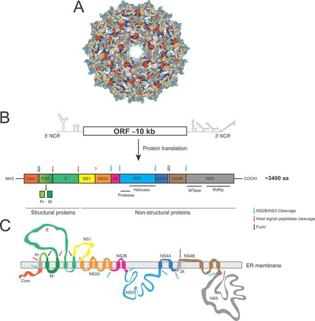
Yellow Fever Virus genome organization (PMID: 29933925)
2.2 Yellow Fever Virus Genome & Classification
The YFV genome is a positive-sense single-stranded RNA of approximately 10.9–11 kb. The genome contains a single open reading frame (ORF) of approximately 10.2 kb, flanked by a short 5′ non-coding region (NCR) and a longer 3′ NCR. The ORF encodes a polyprotein precursor that is post-translationally processed into 10 mature proteins—three structural proteins (capsid protein C, pre-membrane protein prM, and envelope protein E) and seven non-structural proteins (NS1, NS2A, NS2B, NS3, NS4A, NS4B, NS5).
To date, seven genotypes of YFV have been described: five in Africa (West Africa I and II, East Africa, East/Central Africa, Angola) and two in the Americas (South America I and II).
2.3 What Are Yellow Fever Virus Proteins?
Yellow fever virus encodes 10 proteins, including 3 structural proteins and 7 non-structural proteins:
2.3.1 Viral Attachment and Receptor Recognition
Protein | Full name | Main Function |
C | Capsid | Encapsidates the viral genomic RNA, forming the nucleocapsid core |
prM | pre-Membrane | Encapsidates the viral genomic RNA, forming the nucleocapsid core |
E | Envelope | Major surface protein; mediates receptor binding and membrane fusion; primary target of neutralizing antibodies |
2.3.2 Non-structural Proteins
The seven non-structural proteins (NS1, NS2A, NS2B, NS3, NS4A, NS4B, NS5) participate in viral RNA replication, virion morphogenesis, and modulation of host immune responses:
l NS1: secreted protein; can induce vascular endothelial permeability and plays an important role in flavivirus pathogenesis
l NS2B: cofactor for NS3 protease
l NS3: possesses protease and helicase activities; involved in polyprotein processing and viral RNA replication
l NS4B: plays a key role in biogenesis of replication organelles (ROs) and innate immune evasion
l NS5: possesses methyltransferase (5’ cap methylation) and RNA-dependent RNA polymerase (RdRp) activities; core enzyme for viral RNA replication
2.4 Mechanism of Cell Entry and Replication
The yellow fever virus replication cycle resembles that of other flaviviruses and occurs entirely in the host cell cytoplasm.
2.4.1 Viral Attachment and Receptor Recognition
Infection begins with the binding of the viral E protein to host cell surface receptors. LRP8, discovered in 2026, is a functional receptor for YFV. LRP8 expression increases YFV infection in cell lines by promoting viral entry.
2.4.2 Viral Entry
Yellow fever virus enters host cells via receptor-mediated endocytosis. After internalization, endosomal acidification triggers low-pH-induced conformational changes in E protein, mediating fusion of the viral envelope with the endosomal membrane. Following fusion, the viral genomic RNA is released into the host cell cytoplasm.
2.4.3 Viral RNA Replication
Upon release, the viral RNA initiates translation on the endoplasmic reticulum (ER) membrane. The non-structural proteins assemble into replication organelles (ROs) on ER-derived membrane vesicles. NS5 (RdRp) synthesizes a negative-sense RNA intermediate using the positive-sense RNA as template; the negative-sense RNA then serves as template for synthesis of multiple positive-sense genomic RNA copies.
2.4.4 Viral Assembly and Maturation
Newly synthesized viral genomic RNA interacts with structural proteins on the ER membrane and buds into the ER lumen to form immature virions. During transport through the Golgi apparatus, prM protein is cleaved by host furin protease to produce M protein, maturing the virions. Mature virions are finally released via exocytosis.
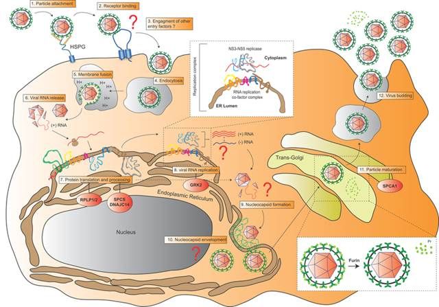
The life cycle of Yellow Fever Virus. (PMID: 29933925)
2.5 Symptoms and Treatment
2.5.1 Clinical Symptoms
The incubation period of yellow fever is typically 3–6 days. Clinical presentation ranges from asymptomatic infection to severe hemorrhagic fever. Approximately 85% of infections are asymptomatic or only mildly symptomatic. Common symptoms in symptomatic patients include:
l Fever
l Headache, myalgia
l Nausea, vomiting
l Jaundice (the name “yellow fever” derives from this)
l Bleeding (gums, nose, gastrointestinal bleeding)
Severe cases may develop hepatitis, disseminated intravascular coagulation (DIC), hemorrhagic manifestations, and multi-organ failure. Case fatality rates in severe patients can reach 50%. In the Americas in 2025, the case fatality rate of yellow fever was 40%–41%.
2.5.2 Antiviral Treatment
Currently, no specific antiviral therapy is approved for yellow fever virus. The WHO 2025 clinical management guidelines recommend a standardized diagnostic and treatment approach.
2.6 Targets for Intervention
2.6.1 Viral Targets
Target | Protein | Biological Role |
E | Envelope protein | Receptor binding & membrane fusion |
NS3 | Protease/helicase | Polyprotein processing, RNA unwinding |
NS4B | Non-structural protein 4B | Replication organelle formation, immune evasion |
NS5 | RNA polymerase (RdRp) | RNA replication |
NS1 | Non-structural protein 1 | Vascular endothelial permeability, immune modulation |
2.6.2 Host Targets
Host Target | Type | Role in Infection |
LRP8 | Receptor | Functional receptor for YFV entry |
LRP4/VLDLR | Receptor | May function as receptors in different tissues |
2.7 Vaccine Types and Development Progress
Yellow fever vaccine is one of the most successful live virus vaccines, widely used since its introduction in the 1930s.
2.7.1 YFV-17D (Live Attenuated Vaccine, Standard Dose)
YFV-17D is the standard live attenuated yellow fever vaccine, in use since the 1930s.
l Efficacy: a single dose is highly effective and provides long-term protection
l Supply constraints: vaccine supply is limited
2.7.2 Low-Dose Yellow Fever Vaccine (Fractional Dosing)
The NIFTY clinical trial (NCT04059471), published in New England Journal of Medicine in 2025, evaluated low-dose yellow fever vaccine in 480 adults in Uganda and Kenya:
l Study design: double-blind, randomized, non-inferiority trial
l Dose groups: standard dose 13,803 IU vs fractional doses of 1000 IU, 500 IU, and 250 IU
l Primary outcome: seroconversion rate at 28 days after vaccination
l Results:
Ø Standard dose seroconversion rate 98%
Ø 500 IU dose differed from standard dose by only 0.01 percentage points (95% CI, -5.0 to 5.1)
Ø Conclusion: yellow fever vaccine doses as low as 500 IU were non-inferior to the standard dose of 13,803 IU for producing seroconversion within 28 days
l Safety: among 111 vaccine-related adverse events, 103 were mild, 7 moderate, and 1 severe
2.7.3 ChAdOx1-Vectored Vaccine
In 2026, the University of Oxford team developed multiple yellow fever virus antigen constructs based on the ChAdOx1 adenovirus vector, using pre-membrane (prM) and envelope (E) proteins—with or without transmembrane domains (TM or ΔTM).
2.7.4 Vaccination Coverage
l African region: by 2024, 26 of 27 high-risk countries had incorporated yellow fever vaccine into routine immunization programs, but average coverage was only 65%
l 2025 Uganda accelerated vaccination campaign: coverage reached 92%
l WHO Eliminate Yellow Fever Epidemics (EYE) strategy: aims to end yellow fever epidemics by 2026
2.8 Drugs
Currently, no specific small-molecule antiviral is approved for yellow fever virus. Several novel antiviral candidates have shown significant progress:
2.8.1 Virus-Targeted Drugs
Target | Drug/candidate | Mechanism | Stage |
NS4B | BDAA (benzodiazepine acetic acid derivative) | Targets NS4B, inhibits viral RNA synthesis and activates dsRNA sensor pathways | Hamster model |
NS5 (RdRp) | 2′-Fluoro-2′-bromouridine monophosphate prodrug | NS5 RdRp inhibition | In vitro biochemical studies |
7-deaza-7-fluoro-2′-C-methyladenosine (DFA) | RdRp inhibition | In vitro | |
Favipiravir | Broad-spectrum antiviral, RdRp inhibition | Hamster model | |
Sofosbuvir | RdRp inhibition | Research setting | |
Multiple targets | Efavirenz (EFV) nanomicelles | Broad-spectrum anti-mosquito-borne flavivirus | Nanoformulation development |
Monoclonal antibody TY014 | Neutralizing antibody | Research setting |
2.8.2 Host-Targeted Drugs
Target | Drug/candidate | Mechanism | Stage |
LRP8 receptor | Soluble LRP8 decoy protein | Blocks YFV infection | Basic research |
Supportive care | Fluid resuscitation, symptomatic treatment | Supportive care | Standard of care |
2.9 Epidemiology
2.9.1 Global Disease Burden
l Approximately 200,000 infections and 30,000 deaths annually
l Severe cases: 84,000–170,000 annually, with 29,000–60,000 deaths
2.9.2 Geographic Distribution
YFV is endemic in 47 countries (34 in Africa and 13 in Central and South America). In 2023, WHO estimated YFV transmission in 34 African countries and 13 Central and South American countries.
2.9.3 2025-2026 Outbreaks
Americas:
l Full year 2025: 7 countries reported 346 confirmed cases and 143 deaths (CFR 41%), a 5.6-fold increase compared with 2024 (61 cases)
l Reporting countries: Bolivia, Venezuela, Brazil, Colombia, Ecuador, Guyana, and Peru
l Brazil: 110 cases, 44 deaths (CFR 40%); Colombia: 60 cases, 24 deaths (CFR 40%)
l In 2025, cases occurred mainly outside the Amazon region, including São Paulo State in Brazil and Tolima Department in Colombia
l First 7 weeks of 2026: 4 countries reported 34 confirmed cases and 15 deaths (CFR 44%)
Africa:
l 2025: 2 outbreaks recorded (Angola and Central African Republic)
l January–May 2026: 3 countries reported 16 confirmed cases (Burkina Faso, Central African Republic, Cameroon)
l An additional 5 countries (Angola, Côte d’Ivoire, Gabon, Ghana, Nigeria) reported suspected cases awaiting investigation
Transmission patterns:
l Main driver: sylvatic transmission cycle—transmission between non-human primates by forest mosquitoes
l The increase in cases in 2025 reflects cyclical reactivation of the sylvatic transmission cycle in the Amazon Basin
l Most human cases had no history of yellow fever vaccination
2.9.4 Transmission Routes
l Mosquito-borne: primarily through bites of Aedes and Haemagogus mosquitoes
l Sylvatic cycle: mosquito → non-human primate → mosquito
l Urban transmission: transmission between humans by Aedes aegypti
2.10 Diagnosis
Diagnosis of yellow fever relies on laboratory testing:
l RT-PCR: detection of viral RNA in blood; detection window ≤14 days; requires specialized laboratory
l Serology (IgM/IgG): attention should be paid to serological cross-reactivity with dengue and other flaviviruses
l Digital PCR (dPCR): enables absolute quantification of ultra-low viral RNA loads in the very early viremic phase
l RT-LAMP: field-deployable detection completed within 1 hour; sensitivity increased 10-fold (LOD 0.29–12 PFU/mL)
l CRISPR-dx (SHERLOCK/DETECTR): ultra-fast sequence-specific detection with low-cost potential
l NGS/metagenomics: for strain characterization and differentiation of wild-type and vaccine-derived viruses
2.11 Prevention
l Vaccination: YFV-17D live attenuated vaccine is the most effective means of preventing yellow fever
l Mosquito control: reduce mosquito breeding sites, use bed nets and repellents
l Personal protection: wear long-sleeved clothing in endemic areas and use repellents
l Traveller precautions: travellers to endemic areas should be vaccinated against yellow fever
l Strengthened surveillance: strengthen monitoring of humans, non-human primates, and mosquito vectors
l Cross-border coordination: strengthen cross-border information sharing and coordinated response
3. Related Products & Services
l Recombinant Antigens & Receptors
l Antibodies
l Detection Kits
l Recombinant Protein Expression Services
l Antibody Development Services
4. Resources
5. References
1. Rice, C. M., Lenches, E. M., Eddy, S. R., et al. (1985). Nucleotide sequence of yellow fever virus: implications for flavivirus gene expression and evolution. Science, 229(4715), 726–733.
2. Chambers, T. J., Hahn, C. S., Galler, R., et al. (1990). Flavivirus genome organization, expression, and replication. Annual Review of Microbiology, 44, 649–688.
3. Mukhopadhyay, S., Kuhn, R. J., Rossmann, M. G. (2005). A structural perspective of the flavivirus life cycle. Nature Reviews Microbiology, 3(1), 13–22.
4. Monath, T. P., Vasconcelos, P. F. C. (2015). Yellow fever. Journal of Clinical Virology, 64, 160–173.
5. Douam, F., Ploss, A. (2018). Yellow Fever Virus: Knowledge Gaps Impeding the Fight Against an Old Foe. Trends in Microbiology, 26(11), 913–928.
6. Shearer, F. M., Moyes, C. L., Pigott, D. M., et al. (2017). Global yellow fever vaccination coverage from 1970 to 2016: a systematic analysis. The Lancet Infectious Diseases, 17(11), 1209–1217.
7. Garske, T., Van Kerkhove, M. D., Yactayo, S., et al. (2014). Yellow fever in Africa: estimating the burden of disease and impact of mass vaccination from outbreak and serological data. PLoS Medicine, 11(5), e1001638.
8. Chong, Z., Hui, S., Qiu, X., et al. (2026). Multiple LDLR family members act as entry receptors for yellow fever virus. Nature, 649(8095), 173–182.
9. Mei, M., Yang, Y., Tan, X., et al. (2026). LRP8 is a functional receptor for yellow fever virus. Nature Microbiology, 11, 1022–1036.
10. Kimathi, D., Juan-Giner, A., Bob, N. S., et al. (2025). Low-Dose Yellow Fever Vaccine in Adults in Africa. The New England journal of medicine, 392(8), 788–797.
11. Schnyder, J. L., de Jong, H. K., Bache, B. E., et al. (2024). Long-term immunity following yellow fever vaccination: a systematic review and meta-analysis. The Lancet. Global health, 12(3), e445–e456.
12. Staples, J. E., Monath, T. P. (2020). Prevention of yellow fever in travellers: an update. The Lancet Infectious Diseases, 20(6), e129–e137.
13. Dubankova, A., Boura, E. (2019). Structure of the yellow fever NS5 protein reveals conserved drug targets shared among flaviviruses. Antiviral research, 169, 104536.
14. Angerami, R. N., Socorro Souza Chaves, T. D., Rodríguez-Morales, A. J. (2025). Yellow fever outbreaks in South America: Current epidemiology, legacies of the recent past and perspectives for the near future. New microbes and new infections, 65, 101580.
15. Weight, A. E., Van Wettere, A. J., Zimmerman, S. M., et al. (2026). BDAA, a yellow fever virus NS4B inhibitor, is effective during therapeutic treatment in a hamster model of yellow fever. Antiviral research, 252, 106464.