
info@mail.ucallm.com
1. Overview
Yersinia enterocolitica is a Gram-negative, facultative anaerobic coccobacillus of the family Yersiniaceae and genus Yersinia, and is the causative agent of yersiniosis, a foodborne zoonotic disease. The bacterium is psychrotrophic, capable of growing at 4℃, thus posing a persistent threat in refrigerated foods.
Y. enterocolitica infection can manifest as acute diarrhoea, mesenteric adenitis, terminal ileitis, and pseudoappendicitis syndrome, with severe cases progressing to sepsis. The bacterium is one of the most common causes of bacterial gastroenteritis in Europe and North America, and in some countries its incidence has surpassed that of Shigella and Salmonella species. Globally, an estimated 117,000 infections occur annually (United States). The species is divided into 6 biotypes (1A, 1B, 2, 3, 4, 5) and over 60 serotypes, with biotype 1B and serotype O:8 being highly pathogenic strains.
2. Background Information
2.1 What is Yersinia enterocolitica?
Y. enterocolitica is a Gram-negative coccobacillus belonging to the family Yersiniaceae and genus Yersinia. The genus includes 26 species, of which three are pathogenic to humans: Y. enterocolitica, Y. pestis, and Y. pseudotuberculosis. Y. enterocolitica is a psychrotrophic bacterium capable of growing and multiplying at refrigeration temperatures, making it a particularly dangerous contaminant in refrigerated foods.
Infection is typically self-limiting, but the organism can continue to be shed in faeces for nearly 3 months after symptoms subside. Post-infectious sequelae include reactive arthritis and erythema nodosum.
2.2 Genome & Classification
The Y. enterocolitica genome is a circular chromosome of approximately 4.6 Mb. Classification is based on biovar and serotype:
Taxonomic Level | Content |
Domain | Bacteria |
Phylum | Pseudomonadota |
Class | Gammaproteobacteria |
Order | Enterobacterales |
Family | Yersiniaceae |
Genus | Yersinia |
Species | Yersinia enterocolitica |
Y. enterocolitica is divided into 6 biotypes, of which biotype 1B is highly pathogenic and carries the high-pathogenicity island (HPI) encoding the yersiniabactin iron transport system. Biotype 1A strains lack known virulence determinants (including the pYV virulence plasmid) but can also cause human infection.
2.3 What Are Y. enterocolitica Virulence Factors?
Y. enterocolitica virulence factors are classified as plasmid-encoded and chromosome-encoded.
2.3.1 Plasmid-Encoded Virulence Factors
All pathogenic Yersinia strains carry an approximately 70-kb virulence plasmid, pYV, which is central to virulence. pYV encodes:
Virulence Factor | Main Function |
YadA | Multifunctional adhesin mediating cell adherence, phagocytosis resistance, serum survival, and collagen binding |
Ysc T3SS | Type III secretion system injecting Yop effectors into host cells |
Yops | Effector proteins suppressing host immune responses |
2.3.2 Chromosome-Encoded Virulence Factors
Virulence Factor | Main Function |
Inv (Invasin) | Outer membrane protein mediating bacterial invasion of intestinal epithelial cells |
Ail | Outer membrane protein mediating adherence and serum resistance |
HPI | High-pathogenicity island encoding yersiniabactin iron transport system |
Ysa T3SS | Chromosome-encoded type III secretion system present only in biotype 1B strains |
2.4 Mechanism of Infection and Pathogenesis
2.4.1 Adhesion and Invasion
Y. enterocolitica infection begins when the bacterium enters the host via the faecal-oral route. Upon reaching the terminal ileum, the bacterium uses invasin (Inv) to invade intestinal epithelial cells and colonizes the lymphoid tissue of Peyer's patches.
2.4.2 Type III Secretion Systems and Yop Effectors
Y. enterocolitica maintains three independent type III secretion systems:
a. Ysc T3SS (plasmid-encoded) : Injects Yop effectors (YopE, YopH, YopP/J, etc.) into host cells, suppressing phagocytosis, inflammatory signalling, and cytoskeletal function.
b. Ysa T3SS (chromosome-encoded) : Present only in biotype 1B strains, functioning during early infection.
c. Flagellar T3SS: Involved in YplA secretion.
2.4.3 Dual Role of YopP In Vivo
A 2025 study published in Medical Microbiology and Immunology revealed an unexpected role for YopP in in vivo infection: contrary to in vitro expectations, a YopP-deficient strain (Ye ΔyopP) increased bacterial dissemination from Peyer's patches to mesenteric lymph nodes and spleen. Mechanistically, YopP regulates the migration of bacteria-carrying dendritic cells from Peyer's patches to mesenteric lymph nodes to limit bacterial dissemination. These findings indicate that YopP plays a complex role in host-pathogen interactions—seemingly benefiting the host while simultaneously facilitating bacterial evasion from early immune clearance..
2.4.4 Biofilm Formation
Y. enterocolitica can form biofilms on abiotic surfaces, an important factor in its persistence in the food chain. A 2026 study published in Food Research International showed that 91.0% of isolates formed biofilms. A 2025 study published in Life found that the virulence plasmid (pYV) modulates glucose-mediated biofilm regulation. Flagellar structure or rotation is crucial for biofilm formation; flagellar mutants greatly reduce biofilm-forming ability.
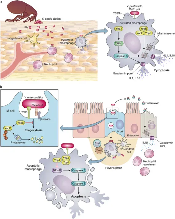
Evolutionary changes impacting adherence and invasion characterize enhancements to the virulence potential of pathogenic Yersiniae. (PMID: 38389313)
2.5 Transmission Routes and Process
Transmission of Y. enterocolitica occurs primarily via the faecal-oral route:
l Foodborne transmission: Ingestion of contaminated food is the most common route, especially raw or undercooked pork and unpasteurized dairy products.
l Waterborne transmission: Drinking contaminated water.
l Contact transmission: Contact with infected animals or humans.
l Transfusion transmission: Via blood transfusion.
Process: Ingestion of contaminated food or water → bacteria reach the terminal ileum → invade intestinal epithelial cells → colonize Peyer's patches → cause local inflammation and diarrhoea → may disseminate to mesenteric lymph nodes (causing pseudoappendicitis) → rarely disseminate to spleen and liver (sepsis).
2.6 Symptoms and Treatment
2.6.1 Clinical Symptoms
Children: Predominantly acute diarrhoea, often with fever and abdominal pain. Infants may present with watery diarrhoea, sometimes bloody.
Older children and adolescents: Characteristic presentation is pseudoappendicitis syndrome, including right lower quadrant pain, fever, and leukocytosis, indistinguishable from appendicitis.
Adults: May present with mesenteric adenitis, terminal ileitis, or reactive arthritis.
Sequelae: Reactive arthritis (HLA-B27-associated) and erythema nodosum.
2.6.2 Treatment
Most infections are self-limiting, and supportive treatment (maintaining water and electrolyte balance) is sufficient.
Indications for antibiotic therapy: Severe symptoms or bloodstream infection.
Drug | Recommendation Level | Considerations |
Fluoroquinolones (ciprofloxacin) | First-line | One of the most effective treatment options |
Third-generation cephalosporins (ceftriaxone) | First-line | Selected based on susceptibility |
Trimethoprim-sulfamethoxazole (TMP-SMX) | Alternative | Most prescribed antimicrobial in New Zealand (49%) |
Doxycycline | Alternative | Based on strain susceptibility |
Resistance: Y. enterocolitica is typically resistant to first-generation cephalosporins and most penicillins (due to beta-lactamase production) but susceptible to TMP-SMX, aminoglycosides, third-generation cephalosporins, fluoroquinolones, and tetracyclines. A 2026 study found that 94% of foodborne and 98% of environmental isolates were resistant to cefazolin, and 9% of strains exhibited multidrug-resistant (MDR) phenotypes.
2.7 Prevention
l Food safety: Thoroughly cook pork (core temperature at least 70°C for 2 minutes); avoid raw or undercooked meat products; separate raw and cooked foods, using separate cutting boards.
l Dairy safety: Consume only pasteurized dairy products.
l Hand hygiene: Thoroughly wash hands before and after preparing food, after contact with animals, and after using the toilet.
l Produce washing: Thoroughly wash vegetables and salads to be eaten raw.
l Kitchen hygiene: Thoroughly clean all cutting boards, countertops, and utensils.
l Child protection: Remove infants and toddlers from the kitchen while preparing pork or chitterlings until all surfaces that came into contact with the food have been cleaned with soap and hot water.
2.8 Targets for Intervention
2.8.1 Pathogen Targets
Target | Protein/Pathway | Biological Function |
Ysc T3SS | Type III secretion system | Yop effector injection |
YadA | Adhesin | Cell adherence, serum resistance |
Inv | Invasin | Intestinal epithelial cell invasion |
HPI | Iron transport system | Iron acquisition |
2.8.2 Host Targets
Target | Type | Role in Infection |
β1 integrin | Receptor | Mediates Yop injection into leukocytes |
Dendritic cells | Immune cells | YopP regulates their migration from Peyer's patches to mesenteric lymph nodes |
2.9 Vaccine Types and Development Progress
Currently, no FDA-licensed human Y. enterocolitica vaccine is available.
2.9.1 Chimeric Vaccine (Reverse Vaccinology Design)
A 2025 study published in Frontiers in Immunology used subtractive proteomics and reverse vaccinology to identify 14 drug targets and 15 vaccine targets from 22 complete genomes. The designed V2 and V4 chimeric vaccine constructs displayed the highest binding affinities with TLR4; immune simulations predicted high immunological profiles, with T-cell epitopes covering 99.26% of the global population.
2.9.2 Multi-Epitope Vaccine (In Silico Design)
A 2024 study published in International Immunopharmacology employed reverse vaccinology to design a multi-epitope vaccine against Y. enterocolitica, providing a promising foundation for experimental validation and vaccine development.
2.10 Drugs
2.10.1 Pathogen-Targeted Drugs
Target | Drug/candidate | Mechanism | Stage |
Outer membrane targets | Chlorogenic acid, hesperidin, neohesperidin, naringin | Molecular docking showed good binding with Y. enterocolitica outer membrane targets | Computational screening |
Multiple targets | Subtractive proteomics-identified targets | 14 non-human homologous essential virulence proteins as drug targets | Computational screening |
2.11 Recent Research
2.11.1 Molecular Epidemiology and Resistance Surveillance
A 2026 study published in Food Research International performed whole-genome sequencing analysis of 50 foodborne and 50 environmental Y. enterocolitica strains. 91.0% of strains formed biofilms, and 50 distinct sequence types (STs) and 70 unique core genome sequence types (cgSTs) were identified, reflecting high genetic diversity. Two pork-derived MDR isolates (YEN2024FY164, YEN2024FY264) exhibited quintuple antibiotic resistance and carried a full complement of key virulence genes.
A 2026 study published in Food Research International analysed the prevalence of Y. enterocolitica in Chinese livestock and poultry products, with an overall prevalence of 9.37%, highest in Southern China (25.00%).
2.11.2 Diagnostic Technology Advances
Dual-gene RAA-lateral flow strip: A 2025-developed dual recombinase-aided amplification (RAA)-lateral flow assay targeting both a pathogenic marker (ail) and a conserved gene (foxA) enables rapid detection of foodborne Y. enterocolitica.
Two-group quadruplex real-time quantitative PCR: A 2025-developed two-group quadruplex RT-qPCR assay for molecular patho-serotyping of Y. enterocolitica.
LAMP-CRISPR one-pot assay: A 2025-developed LAMP-CRISPR one-pot assay enables rapid detection of Y. enterocolitica, overcoming the limitations of the national standard method (time-consuming, not suitable for on-site rapid testing).
β-glucosidase gene PCR: A 2025 study found that the β-glucosidase gene (esculin hydrolysis) of Y. enterocolitica is a suitable PCR target for detecting biotype 1A strains.
2.11.3 Host-Pathogen Interactions
Dual role of YopP: A 2025 study revealed the complex role of YopP in in vivo infection—YopP deficiency increases bacterial dissemination, and YopP limits dissemination by regulating the migration of bacteria-carrying dendritic cells from Peyer's patches to mesenteric lymph nodes.
In-host evolution during chronic infection: A 2025 study published in Nature Communications characterized four Y. enterocolitica clonal isolates from successive bacteraemia episodes that evolved within an elderly patient over 14 years, revealing in-host evolution during chronic infection.
2.11.4 Environmental Survival and Transmission
A 2025 study found that Y. enterocolitica can survive and maintain genomic stability within environmental Acanthamoeba spp..
A 2025 study published in Food Research International analysed multiple points in the food chain (1550 food and 670 environmental samples), with prevalence rates of 25.9% and 23.9% in food and environmental samples, respectively.
3. Related Products & Services
l Recombinant Antigens & Receptors
l Antibodies
l Detection Kits
l Recombinant Protein Expression Services
l Antibody Development Services
4. Resources
5. References
1. Seabaugh, J. A., Anderson, D. M. (2024). Pathogenicity and virulence of Yersinia. Virulence, 15(1), 2316439.
2. Grygiel-Górniak B. (2025). Current Challenges in Yersinia Diagnosis and Treatment. Microorganisms, 13(5), 1133.
3. Fàbrega, A., Vila, J. (2012). Yersinia enterocolitica: pathogenesis, virulence and antimicrobial resistance. Enfermedades infecciosas y microbiologia clinica, 30(1), 24–32.
4. Sabina, Y., Rahman, A., Ray, R. C., Montet, D. (2011). Yersinia enterocolitica: Mode of Transmission, Molecular Insights of Virulence, and Pathogenesis of Infection. Journal of pathogens, 2011, 429069.
5. Cornelis, G. R., Boland, A., Boyd, A. P., et al. (1998). The virulence plasmid of Yersinia, an antihost genome. Microbiology and molecular biology reviews : MMBR, 62(4), 1315–1352.
6. Thomson, N. R., Howard, S., Wren, B. W., et al. (2006). The complete genome sequence and comparative genome analysis of the high pathogenicity Yersinia enterocolitica strain 8081. PLoS genetics, 2(12), e206.
7. Rakin, A., Noelting, C., Schubert, S., et al. (1999). Common and specific characteristics of the high-pathogenicity island of Yersinia enterocolitica. Infection and immunity, 67(10), 5265–5274.
8. Snellings, N. J., Popek, M., Lindler, L. E. (2001). Complete DNA sequence of Yersinia enterocolitica serotype 0:8 low-calcium-response plasmid reveals a new virulence plasmid-associated replicon. Infection and immunity, 69(7), 4627–4638.
9. Kanwal, M., Basheer, A., Bilal, M., et al. (2024). In silico vaccine design for Yersinia enterocolitica: A comprehensive approach to enhanced immunogenicity, efficacy and protection. International immunopharmacology, 143(Pt 1), 113241.
10. Shah, M., Fatima, R., Sarfraz, A., et al. (2025). Screening of novel therapeutic targets and chimeric vaccine construction against antibiotic-resistant Yersinia Enterocolitica. Frontiers in immunology, 16, 1555248.
11. Silva, J. E., Eliçabe, R. J., Davicino, R. C., et al. (2025). YopP modulates bacterial virulence and systemic dissemination in Peyer´s patches during murine Yersinia enterocolitica infection. Medical microbiology and immunology, 214(1), 51. Savin, C., Lê-Bury, P., Guglielmini, J., et al. (2025). In-host evolution of Yersinia enterocolitica during a chronic human infection. Nature communications, 16(1), 5637.
12. Zhou, T., Shen, J., Feng, Y., et al. (2025). Dual-Gene Based Recombinase-Aided Amplification (RAA) - Lateral Flow Strip for the Sensitive On-Site Detection of Yersinia enterocolitica in Food Samples. Journal of AOAC International, 108(4), 620–627.
13. Wang, J., Qin, J., Hu, B., et al. (2025). A two-group quadruplex real-time quantitative PCR assay for molecular patho-serotyping of Yersinia enterocolitica. Letters in applied microbiology, 78(6), ovaf079.
14. Lou, X., Mao, J., Wu, Y., et al. (2026). From foodborne to environmental Yersinia enterocolitica: Comparative insights into antimicrobial resistance and virulence for food safety. Food research international (Ottawa, Ont.), 238, 119442.
15. Fang, Z., Mao, J., Chen, Z., et al. (2026). Deciphering the potential risks of Yersinia enterocolitica across multi-points in food chain: prevalence, biofilm, and cross-stage transmission routes. Food research international (Ottawa, Ont.), 226, 118107.