
info@mail.ucallm.com
From Cardiac Specification to Pathological Remodeling — Empowering Comprehensive Cardiovascular Biology Profiling with Validated Antibody Solutions
Cardiovascular development and remodeling represent highly coordinated biological processes that determine heart formation, vascular maturation, tissue adaptation, and disease progression. From embryonic cardiogenesis to adult cardiac homeostasis, precise regulation of cardiac progenitor differentiation, cardiomyocyte maturation, vascular development, extracellular matrix remodeling, and stress-responsive signaling pathways maintains cardiovascular function.
Following pathological stimuli such as myocardial infarction, pressure overload, hypoxia, and metabolic stress, the heart undergoes dynamic remodeling involving:
- Cardiac progenitor cell specification and lineage commitment
- Cardiomyocyte growth, maturation, and regeneration
- Endothelial differentiation and angiogenic remodeling
- Fibroblast activation and extracellular matrix deposition
- Inflammatory signaling and immune-mediated repair
- Mechanotransduction and stress-responsive pathways
- Epigenetic and transcriptional regulation of cardiac identity
We provide comprehensive antibody solutions for investigating cardiovascular biology—from embryonic heart development and vascular patterning to pathological hypertrophy, fibrosis, myocardial injury repair, and cardiovascular disease mechanisms.
Validated antibody solutions enable researchers to characterize:
- Cardiac developmental transcriptional programs
- Cardiomyocyte identity and maturation states
- Vascular endothelial remodeling and angiogenesis
- Cardiac hypertrophy and fibrosis signaling
- Inflammatory and immune-mediated cardiac repair
- Mechanotransduction and metabolic adaptation
- Molecular mechanisms underlying cardiovascular diseases
Cardiovascular Development & Remodeling Biomarker Antibody Portfolio
——Comprehensive Solutions for Cardiac Development, Vascular Remodeling, and Cardiovascular Disease Discovery
Cardiac development and remodeling are controlled by integrated networks involving transcription factors, receptors, kinases, ion channels, metabolic regulators, and epigenetic modifiers.
Our validated antibody portfolio enables systematic analysis of key molecular pathways involved in:
Research Focus | Representative Biomarkers | Research Applications |
Cardiac Progenitor Specification & Development | NOTCH1, MESP1, ISL1, NKX2.5, GATA4, TBX5, MEF2C | Characterization of cardiac lineage commitment, embryonic cardiogenesis, and progenitor cell differentiation |
Cardiomyocyte Maturation & Structural Remodeling | ACTN2, MYH6, MYH7, TNNT2, TNNI3, DES | Analysis of sarcomere organization, myocardial maturation, and contractile function |
Cardiac Hypertrophy Signaling | TAK1, NFAT, Calcineurin, AKT, mTOR, ERK1/2, p38 MAPK | Investigation of pathological hypertrophy induced by pressure overload and stress signaling |
Cardiac Fibrosis & Extracellular Matrix Remodeling | α-SMA, COL1A1, COL3A1, Fibronectin, TGF-β, SMAD2/3 | Profiling fibroblast activation, matrix deposition, and myocardial fibrosis progression |
Endothelial Development & Angiogenic Remodeling | VEGFR2, CD31, VE-Cadherin, HIF-1α, HIF-2α, FGFR1 | Investigation of vascular formation, endothelial adaptation, and angiogenesis |
Mechanotransduction & Cardiac Stress Response | PIEZO1, YAP/TAZ, Integrin signaling, FAK | Study of mechanical stress sensing and adaptive cardiovascular remodeling |
Cardiac Regeneration & Repair | Ki-67、Aurora B、Phospho-Histone H3 (Ser10), Active β-catenin、Axin2, YAP1,TAZ, Phospho-YAP (Ser127) | Investigation of cardiomyocyte proliferation, regeneration, and tissue repair |
Inflammation & Immune-Mediated Remodeling | NLRP3, NF-κB, IL-1β, TNF-α, TREM2, GSDMD | Analysis of inflammatory activation, immune response, and post-injury remodeling |
Cardiovascular Remodeling Signaling Network
——Integrated Antibody Solutions for Profiling Hypertrophy, Fibrosis, Angiogenesis, and Cardiac Repair Mechanisms
Cardiovascular remodeling is a dynamic, multicellular process driven by coordinated cardiomyocyte stress and hypertrophy, fibroblast activation and ECM remodeling, angiogenesis and vascular adaptation, inflammatory–immune signaling, and cardiac repair/regeneration. Our integrated antibody solutions enable comprehensive profiling of key pathways—including TGF-β/SMAD, YAP/TAZ, MAPK, PI3K–AKT, Wnt/β-catenin, and inflammasome signaling—together with representative biomarkers such as ANP/BNP, α-SMA/FAP/POSTN, collagen, VEGF/VEGFR2, CD31, CD68, NLRP3, and STAT3. By supporting IHC/IF, WB, Flow Cytometry, Multiplex IF, PLA/IP, and spatial or single-cell analyses, this portfolio provides a robust framework to dissect cell–cell crosstalk, mechanical feedback, pathological remodeling, and regenerative responses, accelerating biomarker discovery and therapeutic target validation in cardiovascular disease.
.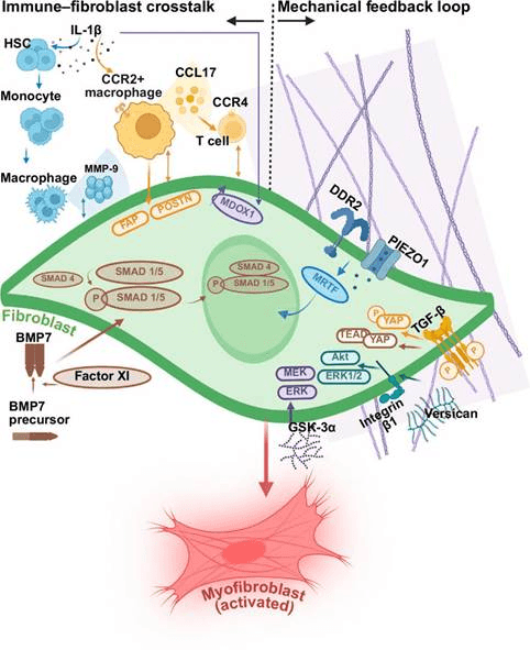
Dual axes of fibrotic regulation: immune crosstalk and mechanical feedback in cardiac fibrosis.(PMID: 40877078)
Featured Cardiovascular Research Panels
——Integrated Antibody Panels for Cardiac Development, Remodeling, and Cardiovascular Disease Discovery
Our cardiovascular research panels provide targeted solutions for dissecting the molecular networks controlling heart development, myocardial adaptation, and pathological remodeling.
Cardiac Development & Progenitor Specification Panel
Profiling Cardiac Lineage Commitment, Differentiation, and Myocardial Formation
Cardiac development requires precise regulation of transcriptional networks controlling progenitor maintenance, differentiation, and chamber formation.
Biological Process | Markers |
Cardiac progenitor identity | NKX2.5, GATA4, TBX5, SALL4 |
Developmental signaling | NOTCH1, Active β-catenin, LEF1, Axin2, BMP4, Phospho-SMAD1/5/9, SMAD4 |
Cardiomyocyte differentiation | TNNT2, ACTN2, MYH6, MYH7 |
Sarcomere maturation | TNNI3, DES, MYL2 |
Cardiac Hypertrophy & Stress Remodeling Panel
Decoding Molecular Mechanisms Driving Pathological Cardiac Growth
Pathological cardiac hypertrophy involves activation of calcium-dependent signaling, inflammatory pathways, and metabolic reprogramming.
Biological Process | Markers |
Calcineurin-NFAT pathway | Calcineurin, NFATc3/NFATc4 |
Stress kinase signaling | TAK1, p38 MAPK, JNK |
Growth signaling | AKT, mTOR, ERK1/2 |
Hypertrophic markers | ANP, BNP, β-MHC/MYH7 |
Cardiac Fibrosis & Matrix Remodeling Panel
Investigating Fibroblast Activation and Extracellular Matrix Remodeling
Fibrotic remodeling contributes to myocardial stiffness, impaired contractility, and progression toward heart failure.
Biological Process | Markers |
Fibroblast activation | α-SMA, PDGFRα |
ECM deposition | COL1A1, COL3A1, Fibronectin |
Fibrotic signaling | TGF-β, p-SMAD2/3 |
Myofibroblast transition | POSTN, FAP |
Vascular Remodeling & Angiogenesis Panel
Mapping Endothelial Adaptation and Cardiovascular Repair
Vascular remodeling requires coordinated regulation of endothelial signaling, hypoxia responses, and angiogenic pathways.
Biological Process | Markers |
Endothelial identity | CD31, VE-Cadherin, EMCN |
Angiogenic signaling | VEGFR2, VEGF-A, FGFR1 |
Hypoxia response | HIF-1α, HIF-2α |
Vascular remodeling | ANGPT1/2, Tie2 |
Cardiac Injury, Inflammation & Repair Panel
Profiling Immune Activation and Post-Injury Remodeling
Following myocardial injury, immune activation coordinates tissue repair but may also contribute to chronic remodeling
Biological Process | Markers |
Inflammasome activation | NLRP3, ASC, Caspase-1 |
Inflammatory signaling | NF-κB, IL-1β, TNF-α |
Macrophage remodeling | TREM2, CD68, F4/80, CD68, CD163 |
Pyroptosis | GSDMD, Cleaved-GSDMD |
Why Researchers Choose Our Cardiovascular Development & Remodeling Antibody Solutions
Validated Cardiac Targets. Integrated Remodeling Panels. Reliable Mechanistic Insights.
Our antibody portfolio supports complete cardiovascular research workflows:
Research Challenge | Our Solution |
How can I characterize cardiac development programs? | Developmental transcription factor and cardiomyocyte identity marker panels |
How can I study pathological cardiac remodeling? | Hypertrophy, fibrosis, and stress signaling antibody solutions |
How can I analyze vascular adaptation? | Endothelial, angiogenesis, and hypoxia-response markers |
How can I investigate myocardial injury responses? | Integrated inflammation, fibrosis, and regeneration panels |
How can I perform spatial cardiac profiling? | IF/IHC-compatible cardiac architecture and remodeling markers |
How can I identify therapeutic targets? | Validated signaling pathway antibodies for mechanism discovery |
Key reference
Hongkun Wang, Zongkuai Yang (2026).TMC6 Is a Novel Therapeutic Target for Pathogenic Cardiac Hypertrophy. Circ Res. 2026 Mar 27;138(7):e327680.
James W McNamara, Ellen B Keen (2026). Alpha protein kinase 3 gene therapy restores heart function in mouse and human models of cardiomyopathy. Nat Cardiovasc Res. 2026 Aug 19.
Silin Liu, Honglin Xu (2026). Myeloid Piezo1 Drives Cardiac Repair Through Orai1-Rac1-Dependent Efferocytosis. Circ Res. 2026 Jul 6;139(2):e328012.
Xue-Xue Zhu, Ao-Yuan Zhang (2026). PGK1 Drives Cardiac Hypertrophy by Regulating the Vimentin/PI3K/Akt Pathway. Circ Res. 2026 Mar 27;138(7):e327173.
Yingrui Li, Jianlin Du (2024). The molecular mechanisms of cardiac development and related diseases. Signal Transduct Target Ther. 2024 Dec 23;9(1):368.