
info@mail.ucallm.com
From Immune Regulation to Therapeutic Mechanism Discovery — Empowering Comprehensive Immune Checkpoint Profiling with Validated Antibody Solutions
Immune checkpoints are critical regulators of immune homeostasis, controlling the balance between immune activation, immune tolerance, and immune-mediated tissue damage. These regulatory pathways are dynamically controlled through a complex network of co-inhibitory and co-stimulatory receptors, which modulate T-cell activation, exhaustion, differentiation, survival, and anti-tumor immune responses.
In cancer, chronic infection, autoimmune disorders, and immunotherapy-related studies, dysregulated checkpoint signaling contributes to:
T-cell exhaustion and impaired immune surveillance
Tumor immune escape and immunosuppressive microenvironment formation
Altered antigen-presenting cell (APC) activation
Dysfunctional NK-cell and cytotoxic lymphocyte responses
Resistance mechanisms to immune checkpoint blockade therapies
Comprehensive characterization of immune checkpoint pathways requires integrated profiling of receptor expression, downstream signaling, immune cell states, and tumor–immune interactions.
Our validated antibody portfolio enables researchers to investigate:
Immune checkpoint receptor expression and regulation
T-cell activation, exhaustion, and memory formation
Tumor immune microenvironment remodeling
Immune escape mechanisms
Checkpoint inhibitor response and resistance pathways
Combination immunotherapy strategies
Immune Checkpoint Receptor Antibody Portfolio
—Comprehensive Antibody Solutions for Mapping Immune Suppression, Immune Activation, and Therapeutic Response
Immune checkpoint signaling is governed by a dynamic balance between inhibitory and stimulatory receptor pathways. Profiling these receptor networks provides critical insights into immune cell function, disease progression, and therapeutic intervention strategies.
1. Co-stimulatory Immune Checkpoint Receptors
—Characterizing Immune Activation, T-cell Expansion, and Anti-tumor Effector Function
Target | Full Name | Research Association |
CD28 | Cluster of Differentiation 28 | Essential co-stimulatory receptor promoting T-cell activation, proliferation, and survival |
ICOS | Inducible T-cell Costimulator | Regulates Tfh/Tfr differentiation, T-cell activation, and T–B cell communication |
OX40 (CD134) | Tumor Necrosis Factor Receptor Superfamily Member 4 | Promotes T-cell survival, expansion, and memory formation |
4-1BB (CD137) | Tumor Necrosis Factor Receptor Superfamily Member 9 | Enhances cytotoxic T-cell activity, persistence, and immune memory |
GITR | Glucocorticoid-Induced TNFR-Related Protein | Regulates effector T-cell activation and Treg-mediated immune suppression |
CD27 | Tumor Necrosis Factor Receptor Superfamily Member 7 | Supports T-cell activation, survival, and memory differentiation |
CD40 | CD40 Molecule | Activates antigen-presenting cells and regulates B-cell responses and adaptive immunity |
CD226 (DNAM-1) | DNAX Accessory Molecule 1 | Promotes T/NK-cell activation and cytotoxic immune responses |
2. Co-inhibitory Immune Checkpoint Receptors
—Profiling Immune Suppression, T-cell Exhaustion, and Tumor Immune Escape Mechanisms
Target | Full Name | Research Association |
PD-1 | Programmed Cell Death Protein 1 | Central regulator of T-cell exhaustion, immune tolerance, and suppression of anti-tumor immune responses |
CTLA-4 | Cytotoxic T-Lymphocyte–Associated Protein 4 | Competitive inhibition of CD28-mediated co-stimulation through CD80/CD86 binding; regulates early T-cell activation |
LAG-3 | Lymphocyte Activation Gene 3 | Associated with exhausted T-cell phenotypes, impaired TCR signaling, and immune suppression |
TIM-3 | T-cell Immunoglobulin and Mucin-Domain Containing 3 | Regulates T-cell dysfunction, immune tolerance, and chronic inflammatory responses |
TIGIT | T-cell Immunoreceptor with Ig and ITIM Domains | Controls T/NK-cell inhibition, cytotoxic function, and tumor immune escape |
VISTA | V-domain Ig Suppressor of T-cell Activation | Mediates myeloid-associated immune suppression and regulation of T-cell activation |
BTLA | B and T Lymphocyte Attenuator | Maintains immune tolerance through lymphocyte inhibitory signaling |
CD96 | CD96 / T-cell Surface Protein | Regulates NK-cell and T-cell activity through checkpoint signaling networks |
PVRIG | PVR-related Ig Domain-containing Protein | Modulates NK/T-cell responses through CD155/CD112-associated checkpoint pathways |
B7-H3 | CD276 Molecule | Suppresses T-cell activation, promotes tumor immune evasion |
B7-H4 | VTCN1 / V-set domain-containing T-cell activation inhibitor 1 | Inhibits T-cell proliferation, cytokine production, and anti-tumor immunity |
LILRB2/ILT4 | Leukocyte Immunoglobulin Like Receptor B2 | Myeloid checkpoint receptor that binds HLA-G and classical MHC class I, suppressing macrophage and dendritic cell activation, |
Immune Checkpoint Molecular Network
—Integrated Antibody Solutions for Decoding Immune Regulation, Cellular Crosstalk, and Immunotherapy Response Mechanisms
Immune checkpoint signaling represents a highly interconnected regulatory network that coordinates T-cell activation, immune tolerance, tumor immune escape, and therapeutic response. Rather than functioning as isolated pathways, checkpoint receptors integrate signals from co-inhibitory receptors, co-stimulatory receptors, antigen-presenting cells, tumor cells, and the surrounding immune microenvironment.
Comprehensive profiling of these molecular networks enables researchers to understand:
Immune activation versus suppression balance
T-cell exhaustion and functional decline
Tumor immune evasion mechanisms
Immune checkpoint blockade sensitivity and resistance
Cellular interactions within the tumor microenvironment
Our validated antibody portfolio supports systematic investigation of major immune checkpoint signaling networks across cancer immunology and translational immunotherapy research.
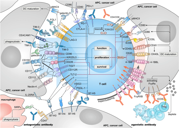
Antagonistic and agonistic antibodies act on inhibitory immune checkpoints and co-stimulatory molecules respectively to promote anti-tumoral T cell functions.(PMID: 38773064)
Featured Immune Checkpoint Research Panels
—Integrated Antibody Solutions for Immunotherapy Discovery and Tumor Immune Profiling
Immune checkpoint pathways orchestrate the balance between immune activation, immune tolerance, and tumor immune evasion.
Our validated antibody panels enable comprehensive profiling of checkpoint receptor networks, immune cell states, and therapeutic response mechanisms across cancer immunology and translational research.
n Immune Exhaustion & Checkpoint Blockade Response Panel—Profiling exhausted T-cell states and immunotherapy responsiveness
Key Markers
PD-1 | CTLA-4 | LAG-3 | TIM-3 | TIGIT |
CD39 | TOX | Eomes | T-bet | Granzyme B |
n Tumor Immune Microenvironment Panel—Mapping immune cell interactions and checkpoint regulation within tumors
Key Markers
T Cells | CD3 | CD4 | CD8 | FOXP3 | Granzyme B |
Myeloid Cells | CD68 | CD163 | CD11b | HLA-DR | CSF1R |
Checkpoint Molecules | PD-L1 | PD-1 | TIGIT | VISTA | LAG-3 |
n T-cell Activation & Co-stimulation Panel—Investigating immune activation states and effector function
Key Markers
CD28 | ICOS | OX40 | 4-1BB | CD27 | CD226 |
CD69 | CD25 | Ki-67 | Granzyme B | IFN-γ | FOXP3 |
n Myeloid Immunoregulation & Immune Suppression Panel—Profiling Macrophage Polarization, Myeloid Checkpoint Signaling, and Tumor Immune Suppression
Key Markers
CD68 | CD163 | CD206 | ARG1 |
iNOS | CSF1R | VISTA | SIRPα |
Why Researchers Choose Our Immune Checkpoint Antibody Solutions
Validated Targets. Comprehensive Immune Pathway Coverage. Reliable Immunotherapy Insights.
Our antibody portfolio supports integrated immune checkpoint research workflows:
Research Challenge | Antibody Solution |
How can I analyze immune checkpoint activation? | PD-1, CTLA-4, LAG-3, TIM-3, TIGIT receptor profiling |
How can I characterize T-cell exhaustion? | Exhaustion marker panels including PD-1, TOX, TIM-3, LAG-3 |
How can I evaluate immunotherapy response? | Checkpoint receptor and immune activation profiling |
How can I study tumor immune microenvironment? | Multiplex IF/IHC-compatible immune marker panels |
How can I analyze immune cell interactions? | T-cell, NK-cell, macrophage, and APC marker solutions |
How can I validate therapeutic targets? | Validated antibodies compatible with WB, IHC, ICC/IF, Flow Cytometry |
Key reference
1. Luo Y, Yuan Y, Liu D, Peng H, Shen L, Chen Y. Targeting novel immune checkpoints in the B7-H family: advancing cancer immunotherapy from bench to bedside. Trends Cancer. 2025 Jun;11(6):540-559.
2. Kraehenbuehl L, Weng CH, Eghbali S, Wolchok JD, Merghoub T. Enhancing immunotherapy in cancer by targeting emerging immunomodulatory pathways. Nat Rev Clin Oncol. 2022 Jan;19(1):37-50.
3. Merhi M, Ahmad F, Taib N, Inchakalody V, Uddin S, Shablak A, Dermime S. The complex network of transcription factors, immune checkpoint inhibitors and stemness features in colorectal cancer: A recent update. Semin Cancer Biol. 2023 Feb;89:1-17.
4. Sathe A, Ayala C, Bai X, Grimes SM, Lee B, Kin C, Shelton A, Poultsides G, Ji HP. GITR and TIGIT immunotherapy provokes divergent multicellular responses in the tumor microenvironment of gastrointestinal cancers. Genome Med. 2023 Nov 26;15(1):100.
5. Ayata P, Crowley JM, Challman MF, Sahasrabuddhe V, et.al. Lymphoid gene expression supports neuroprotective microglia function. Nature. 2025 Dec;648(8092):157-165.