
info@mail.ucallm.com
Natural exosomes possess favorable biocompatibility, low immunogenicity and innate cell-targeting capacity, making them novel biological delivery carriers. Nevertheless, the abundance of functional cargo in native exosomes is limited, and exosomes from different sources exhibit heterogeneity in molecular composition and biological effects. Accordingly, research focus has gradually expanded from functional characterization of native exosomes to the development of engineered exosomes in recent years. By modulating exosome biogenesis, performing surface modification and adopting cargo loading strategies, efficient delivery of specific functional molecules can be achieved.
The engineered exosome platform mainly includes three technical directions: cargo loading, surface engineering and targeting modification. Among them, cargo loading serves as the core technology for utilizing exosomes as therapeutic delivery carriers. Target RNAs, proteins or small-molecule drugs are artificially loaded into exosomes, enabling stable in vivo transport and action on specific cells.
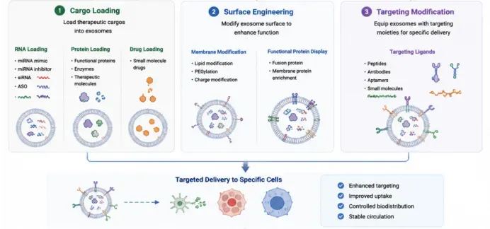
Construction and targeted delivery of engineered exosomes. Functional upgrading of engineered exosomes is realized via cargo loading, surface engineering and targeting modification: encapsulation of nucleic acids, proteins and small‑molecule drugs; optimization of vesicle performance through membrane modification and functional protein display; conjugation of targeting ligands to achieve specific recognition. Modified exosomes can target specific cells, enhance targeting capability and cellular uptake, regulate in vivo distribution and sustain long‑term circulation.
1 Exosome Cargo Loading
Exosomes naturally carry a variety of bioactive molecules including mRNA, miRNA, proteins and lipids. However, higher loading efficiency of specific functional molecules is required for disease therapy and precise regulation. We therefore provide multiple engineered loading strategies, namely endogenous loading and exogenous loading.
Endogenous loading strategies mainly adopt genetic engineering to modify donor cells so that they express target RNA or protein molecules. The cellular intrinsic exosome biogenesis pathway is harnessed to selectively package target cargo into newly formed exosomes. For example, donor cells can be transfected to express specific miRNA, siRNA or fusion proteins, yielding exosomes enriched with target molecules.
Exogenous loading strategies directly introduce functional molecules into isolated exosomes by physical or chemical approaches, including electroporation, sonication, extrusion, membrane permeabilization and lipid fusion. Such methods feature flexible operation and compatibility with diverse cargo types, and are widely applied in the research of nucleic acid drug and protein delivery.
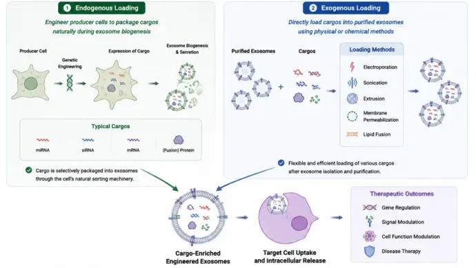
Technical routes for cargo loading of engineered exosomes: endogenous loading versus exogenous loading. Two major loading strategies are available for therapeutic cargo loading into exosomes. Endogenous loading genetically modifies donor cells. During natural biogenesis and secretion of vesicles, biomolecules such as nucleic acids and fusion proteins are packaged via cellular sorting pathways. Exogenous loading directly loads various cargos into purified exosomes using physical and chemical methods including electroporation, sonication and membrane permeabilization. Engineered exosomes enriched with cargo prepared by these two approaches release active molecules after entering target cells, regulating genes and cellular signals to achieve disease treatment.
Owing to their innate capacity to protect nucleic acids and facilitate cellular uptake, exosomes serve as an ideal platform for delivering nucleic acid therapeutics. Currently, engineered exosomes are mainly used for delivery of miRNA mimics, miRNA inhibitors, siRNA and antisense oligonucleotides (ASOs).
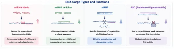
Types and working mechanisms of exosomal RNA cargo. A variety of RNA molecules can be used as therapeutic exosome cargo to achieve precise gene regulation. miRNA mimics replenish downregulated miRNAs in vivo to restore cellular homeostasis; miRNA inhibitors antagonize overabundant miRNAs and de-repress the expression of target genes; siRNAs specifically silence disease-causing genes through the RNA interference pathway; antisense oligonucleotides (ASOs) bind target RNAs, regulate splicing, suppress translation or trigger degradation of target RNAs, meeting diverse requirements for gene intervention.
Among them, miRNA mimics are applied to supplement functional miRNAs lost under disease conditions, whereas miRNA inhibitors and siRNAs can specifically suppress the expression of abnormally activated target genes. Antisense oligonucleotides (ASOs) bind complementarily to target RNAs to induce RNA degradation or modulate translation. For instance, engineered exosomes can deliver inhibitory siRNA targeting mutant KRAS, effectively reducing mutant KRAS expression and restraining tumor growth in pancreatic cancer models.
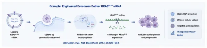
Application Case: Delivery of KRASG12D siRNA by Engineered Exosomes. This case represents a classic study on exosome-mediated nucleic acid delivery. siRNA targeting the KRASG12D mutation is loaded into engineered exosomes. The exosomes can be efficiently internalized by pancreatic cancer cells and release siRNA intracellularly, specifically silencing the mutant KRAS gene and arresting tumor proliferation. The exosome carrier protects nucleic acids, enables efficient cellular uptake and precise gene regulation, and exerts anti-tumor efficacy in vivo. Reference: Kamerkar et al., Nat. Biotechnol. 2017; 35:585–594.
Products: In response to the challenge of insufficient efficiency in exosome nucleic acid delivery in recent years, we offer multiple high-efficiency cargo loading technologies. Among them, the ExoLoad® platform optimizes the exosome loading workflow to achieve high-efficiency encapsulation of small nucleic acids including miRNA mimics, miRNA inhibitors and siRNAs. This technology improves the proportion of functional nucleic acids incorporated into exosomes while preserving exosome membrane integrity and biological activity. Engineered exosomes generated by this platform serve as valuable tools for investigating gene regulation, disease models and nucleic acid drug delivery.
Product Category | Product Name | Catalog No. | Specification | Core Application |
EV Nucleic Acid Loading | ExoLoad® Nucleic Acid Loading Kit | ELSR-06-02 | 3E11 × 4T | For efficient loading of small nucleic acids such as miRNA mimics, miRNA inhibitors and siRNAs into EVs/exosomes for gene regulation and functional studies |
EV Nucleic Acid Loading | ExoLoad® Nucleic Acid Loading Kit | ELSR-06-03 | 1.5E12 × 6T | Designed for large-scale EV nucleic acid loading, suitable for engineered exosomes and nucleic acid delivery research |
2 Surface Engineering
Natural exosomes possess favorable biocompatibility and low immunogenicity, together with innate capacity for intercellular communication. However, their in vivo trafficking is affected by donor cell characteristics, tissue barriers and non-specific clearance mechanisms, which makes it difficult for exosomes to achieve highly precise tissue targeting and cell-selective delivery. Therefore, with the advancing therapeutic applications of exosomes, researchers have developed surface engineering technologies for exosomes. By modifying the molecular composition and recognition properties of the exosomal membrane surface, these approaches enhance targeting performance, tissue localization and cellular uptake efficiency, transforming exosomes from natural information carriers into programmable precise delivery platforms.
Exosome surface modification relies largely on the native lipid bilayer and membrane protein localization features of exosomes. Strategies including lipid anchoring, membrane protein engineering, targeting peptide display, and antibody/ligand conjugation are used to introduce recognition molecules onto the exosome surface. Such engineering endows exosomes with new biological properties, enabling them to more effectively recognize receptors on target cell surfaces and promote exosome accumulation in specific tissues.
2.1 Lipid Anchoring
Lipid anchoring is a surface functionalization method based on the lipid structure of exosomal membranes. Since exosomal membranes are mainly composed of cholesterol, phospholipids, sphingomyelin and other lipids with strong lipid-fusion capacity, lipid-linked structures with membrane-inserting properties can be used to stably anchor functional molecules onto the exosome surface.
Commonly used lipid anchors include DSPE‑PEG, PEG‑lipid and other lipid-linked moieties. Targeting peptides, antibody fragments, fluorescent probes or functional proteins can be conjugated to lipid structures, which insert into the exosomal lipid bilayer via hydrophobic interactions to achieve surface functionalization. Compared with genetic engineering strategies, lipid anchoring does not alter the genetic background of exosome donor cells, featuring simple operation, broad applicability and high modification efficiency.
Lipid-anchor modification not only strengthens the interaction between exosomes and target cells, but also improves exosome stability in vivo. For example, PEGylated lipid modification reduces rapid clearance of exosomes by the mononuclear phagocyte system, prolongs blood circulation time and boosts in vivo delivery efficiency. In addition, conjugation of tissue-specific targeting molecules onto exosomes via lipid anchoring facilitates exosome enrichment in target tissues, offering new technical solutions for precise drug delivery.
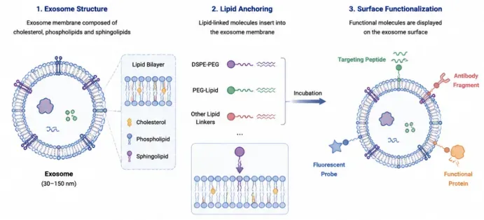
Basic structure of exosomes and surface functional modification via lipid anchoring. Exosomes are 30–150 nm bilayer membrane vesicles, and their membrane components include cholesterol, phospholipids and sphingolipids. Lipid anchoring is a straightforward in vitro modification method: lipid derivatives such as DSPE‑PEG spontaneously insert into the exosomal membrane upon incubation. Using these lipids as linkers, targeting peptides, antibody fragments, fluorescent probes, functional proteins and other molecules can be conjugated to the vesicle surface for rapid targeted engineering and labeling of exosomes, with no need for cellular genetic modification.
Products:
Product | Catalog No. | Type |
ExoBrooch®-RVG Lipid Anchoring Kit | EA-06-5 | Targeted Lipid Anchoring |
ExoBrooch®-iRGD Lipid Anchoring Kit | EA-08-5 | Targeted Lipid Anchoring |
ExoBrooch®-IMTP Lipid Anchoring Kit | EA-11-5 | Targeted Lipid Anchoring |
ExoBrooch® Targeting Peptide Lipid Anchoring Custom Service | — | Custom Service |
Learn about RVG Lipid-Achor Kit-A Tool for Neural-Targeted Exosome Engineering)
2.2 Membrane Protein Engineering
Membrane protein engineering is one of the most widely adopted approaches in exosome surface engineering research. This strategy leverages membrane proteins naturally enriched on exosomes as molecular display scaffolds. Through genetic engineering, target functional molecules are fused and expressed with exosome membrane-localized proteins, which then localize to the vesicle surface during exosome biogenesis.
Membrane proteins commonly used for exosome surface display include Lamp2b, CD63, CD81 and CD9. Among them, Lamp2b serves as the most widely used scaffold for exosome engineering and display owing to its stable exosomal membrane localization and compatibility for fusion expression with various targeting peptides. By constructing "functional molecule-Lamp2b" fusion proteins, researchers can enable specific localization of targeting domains or functional proteins on the exosome surface, thereby enhancing recognition between exosomes and recipient cells.
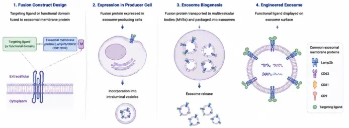
Workflow for preparing engineered exosomes via fusion protein construction. Targeting ligands or functional fragments are linked to classic exosomal membrane proteins including Lamp2b, CD63, CD81 and CD9. After plasmid transfection of donor cells, the cells express fusion proteins, which are sorted into intraluminal vesicles of multivesicular bodies. Multivesicular bodies fuse with the cell membrane and release exosomes. Consequently, targeting ligands are stably displayed on the exosomal membrane surface to achieve targeted functional modification of exosomes.
The landmark study published by Alvarez-Erviti et al. in Nature Biotechnology first demonstrated the value of the membrane protein fusion strategy for targeted exosome delivery. Using dendritic cell-derived exosomes, researchers genetically fused the neuron-targeting peptide RVG (rabies virus glycoprotein-derived peptide) with Lamp2b, enabling RVG display on the exosomal membrane surface. Upon intravenous injection, these engineered exosomes efficiently entered the mouse brain and delivered BACE1 siRNA to reduce BACE1 expression in brain tissue, laying an important foundation for nucleic acid drug delivery in neurological diseases. This study proved that membrane protein fusion expression can endow exosomes with new tissue-targeting capacity, converting native cellular communication carriers into programmable therapeutic delivery platforms.
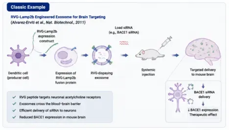
Classic Literature Case — RVG-Lamp2b engineered exosomes for brain-targeted drug delivery. This landmark study lies in the field of targeted delivery of engineered exosomes. Dendritic cells are selected as vesicle-producing cells. The RVG-Lamp2b fusion construct is transfected into cells, enabling secreted exosomes to display the RVG targeting peptide on their surface. BACE1 siRNA is then loaded into exosomes. After systemic administration, RVG recognizes neuronal acetylcholine receptors and assists exosomes in crossing the blood-brain barrier to deliver siRNA to brain neurons and suppress BACE1 gene expression. This work validates the feasibility of exosomes for central nervous system nucleic acid drug delivery.
2.3 Targeting Peptide Modification
Targeting peptide modification is an engineering strategy that employs short peptide molecules to specifically recognize cell-surface receptors and enhance exosome targeting capacity. Given that many cell types exhibit distinct expression patterns of membrane receptors, displaying corresponding targeting peptides on exosome surfaces promotes preferential binding of exosomes to target cells and improves cargo delivery efficiency.
Widely investigated targeting peptides include the neuron-targeting RVG peptide, tumor-targeting RGD peptide and tumor-penetrating iRGD peptide. The RVG peptide recognizes nicotinic acetylcholine receptors on neurons and is therefore extensively used to construct brain-targeted exosomes. The RGD peptide binds αvβ3 integrin receptors expressed on tumor cells and vascular endothelial cells, and is commonly adopted for tumor-targeted delivery research. In oncology studies, targeting peptide modification increases exosome accumulation within tumor tissues and boosts delivery efficiency of anti-tumor drugs, siRNAs and immunomodulatory molecules. For instance, after introducing RGD sequences onto the exosomal membrane, engineered exosomes display enhanced binding to tumor cells with high αvβ3 expression and facilitate cellular uptake, offering a new delivery strategy for precision therapy of solid tumors.
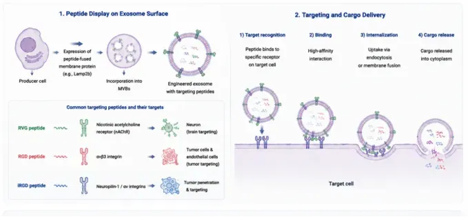
Targeting peptide-modified engineered exosomes: surface construction and targeted delivery mechanisms. Using genetic fusion technology, targeting peptides are fused and co-expressed with the Lamp2b exosomal membrane protein in donor cells, displaying the targeting peptides on the exosome surface during vesicle biogenesis. RVG, RGD and iRGD are the most commonly used targeting peptides in research, acting on neurons and tumor cells, respectively. The modified exosomes achieve tissue/cell-specific delivery through receptor recognition, specific binding, endocytic internalization and eventual release of internal cargo.
In addition, Hoshino et al. reported in Nature that the integrin repertoire on the natural exosome surface determines organotropism. Different integrin expression patterns influence interactions between exosomes and specific tissue cells. For example, αvβ5 integrin promotes exosome uptake by hepatic Kupffer cells, whereas α6β4 and α6β1 integrins are associated with lung tissue localization. This finding further demonstrates that exosomal surface molecules play key roles in governing tissue localization and intercellular communication.
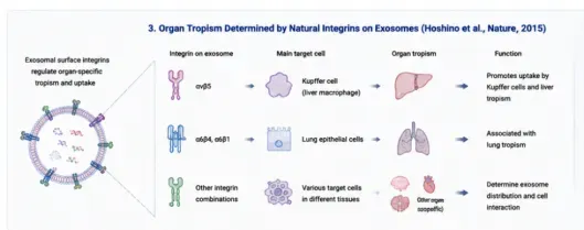
Classic Study: Native Exosomal Integrins Determine Organotropism (Hoshino, Nature, 2015). Integrin proteins carried by exosomes themselves are key molecules governing tissue-targeted distribution in vivo. Integrin αvβ5 can be recognized by hepatic Kupffer cells and drives exosome accumulation in the liver; integrins α6β4 and α6β1 mediate exosome targeting to pulmonary epithelial cells and confer lung tropism. Distinct integrin combinations can guide exosome colonization in different organs in vivo. This principle provides an important theoretical basis for the targeted design of both native and engineered exosomes.
Products:
Product Name | Catalog No. | Specification | Application Scenario | Features |
ExoBrooch®-RVG | EA-06-5 | F5000 | Target receptor: nAChRα7; Targeting type: Nervous system | Generally high yield, sufficient targeted exosomes can be obtained in one batch for in vivo experiments. Two-step procedure with only 30 min hands-on time for targeting peptide conjugation to exosomes and maximum single-batch throughput. Effectively addresses technical challenges including difficulties in modifying non-immortalized cells and various pre-modification issues. Positive rate can reach 70%–80% for the 293F exosome kit. |
ExoBrooch®-iRGD | EA-08-5 | F5000 | Target receptor: Integrin; Targeting type: Pan-cancer endothelial cells and blood vessels in breast cancer, osteosarcoma, ovarian cancer, lung cancer, colon cancer, melanoma, pancreatic cancer, liver cancer, etc. | Suitable for multiple studies on tumor-targeted exosomes. |
ExoBrooch®-IMTP | EA-11-5 | F5000 | Target receptor: Ischemic myocardial tissue; Targeting type: Cardiovascular diseases | For research on tissue-targeted exosomes related to cardiovascular diseases. |
Targeting Peptide Lipid Custom Service | / | 5 mg | Exosome input (divided into 5 runs): 7.3E+12 particles. Custom lipid anchors with known targeting peptide sequences as requested by customers; peptide length less than 30 amino acids | 5000 μg; total exosome input (divided into 5 runs): 7.3E+12 particles. |
Products:
Product Name | Catalog No. | Specification | Application |
ExoCoupl® Exosome Surface Modification Bioorthogonal Conjugation Kit | EC-01-02 | 6T | Used for surface loading of small molecules, small nucleic acids, peptides, etc. onto EVs. Customers need to custom-synthesize N3-modified small molecules, small nucleic acids or peptides.Supports 6 bioorthogonal conjugation reactions, processing 500 μl × 6 = 3 ml exosome samples. Recommended exosome concentration: >1E+11 particles/ml or 0.5–1 μg/μl. |
Learn About Our In Vivo Targeting Products
References
[1] Valadi H, Ekström K, Bossios A, et al. Exosome-mediated transfer of mRNAs and microRNAs is a novel mechanism of genetic exchange between cells. Nat Cell Biol. 2007;9(6):654–659. doi:10.1038/ncb1596.
[2] Kalluri R, LeBleu VS. The biology, function, and biomedical applications of exosomes. Science. 2020;367(6478):eaau6977. doi:10.1126/science.aau6977.
[3] van Niel G, D’Angelo G, Raposo G. Shedding light on the cell biology of extracellular vesicles. Nat Rev Mol Cell Biol. 2018;19(4):213–228. doi:10.1038/nrm.2017.125.
[4] Théry C, Witwer KW, Aikawa E, et al. Minimal information for studies of extracellular vesicles 2018 (MISEV2018): a position statement of the International Society for Extracellular Vesicles and update of the MISEV2014 guidelines. J Extracell Vesicles. 2018;7(1):1535750. doi:10.1080/20013078.2018.1535750.
[5] Villarroya-Beltri C, Gutiérrez-Vázquez C, Sánchez-Cabo F, et al. Sumoylated hnRNPA2B1 controls the sorting of miRNAs into exosomes through binding to specific motifs. Nat Commun. 2013;4:2980. doi:10.1038/ncomms3980.
[6] Alvarez-Erviti L, Seow Y, Yin H, et al. Delivery of siRNA to the mouse brain by systemic injection of targeted exosomes. Nat Biotechnol. 2011;29(4):341–345. doi:10.1038/nbt.1807.
[7] Kamerkar S, LeBleu VS, Sugimoto H, et al. Exosomes facilitate therapeutic targeting of oncogenic KRAS in pancreatic cancer. Nature. 2017;546(7659):498–503. doi:10.1038/nature22341.
[8] Hoshino A, Costa-Silva B, Shen TL, et al. Tumour exosome integrins determine organotropic metastasis. Nature. 2015;527(7578):329–335. doi:10.1038/nature15756.
[9] Chen G, Huang AC, Zhang W, et al. Exosomal PD-L1 contributes to immunosuppression and is associated with anti-PD-1 response. Nature. 2018;560(7718):382–386. doi:10.1038/s41586-018-0392-8.
[10] Vader P, Mol EA, Pasterkamp G, Schiffelers RM. Extracellular vesicles for drug delivery. Adv Drug Deliv Rev. 2016;106:148–156. doi:10.1016/j.addr.2016.02.006.
[11] Kooijmans SAA, Aleza CG, Roffler SR, et al. Display of GPI-anchored anti-EGFR nanobodies on extracellular vesicles promotes tumour cell targeting. J Extracell Vesicles. 2016;5:31053. doi:10.3402/jev.v5.31053.