
info@mail.ucallm.com
From Immune Profiling to Precision Cellular Characterization — Empowering Advanced Immuno-Oncology, Cancer Stem Cell, and Translational Research with Validated CD Antibody Solutions
The Cluster of Differentiation (CD) marker system represents one of the most widely adopted molecular frameworks for cellular identification, immune phenotyping, and tumor microenvironment analysis. CD molecules function as highly informative cellular “identity markers,” regulating diverse biological processes including immune activation, cell adhesion, migration, differentiation, signal transduction, and stemness maintenance.
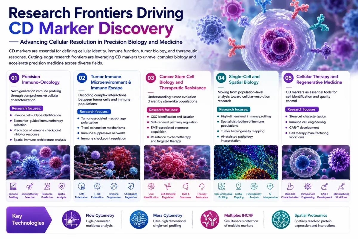
We support next-generation CD marker research by enabling high-resolution characterization of immune cells, tumor ecosystems, stem-like populations, and therapeutic cell products. Validated CD marker solutions empower precision immuno-oncology studies, including immune profiling, checkpoint regulation, tumor immune escape, and biomarker-guided therapy development. Combined with flow cytometry, mass cytometry, multiplex IHC/IF, and spatial proteomics technologies, CD-based profiling reveals cellular heterogeneity, immune architecture, and functional states within complex tissues. These approaches accelerate discoveries in cancer biology, immunotherapy, regenerative medicine, and advanced cellular therapy development.
CD Marker Antibody Portfolio
——Comprehensive Solutions for Immune Profiling, Tumor Characterization, and Stem Cell Research
Accurate cellular identification requires integrated analysis of lineage markers, activation markers, immune regulatory molecules, tumor-associated CD proteins, and stemness-associated biomarkers.
Our CD antibody portfolio covers major research categories to support standardized workflows across immunology, oncology, neuroscience, and regenerative medicine.
CD Marker Category | Representative Markers | Research Applications | Biological Significance |
Immune Lineage Identification Markers | CD3, CD4, CD8, CD19, CD20, CD14, CD68 | Immune cell population profiling and tissue immune mapping | Define immune cell origin and composition within tissues |
Immune Activation & Functional Markers | CD25, CD38, CD69, CD80, CD86 | Immune activation monitoring and functional characterization | Reflect immune cell activation status and immune response |
Immune Checkpoint & Exhaustion Markers | PD-1 (CD279), PD-L1 (CD274), CTLA-4 (CD152), LAG-3, TIM-3 | Immunotherapy response studies and immune suppression analysis | Reveal mechanisms of immune escape and T-cell dysfunction |
Cancer Stem Cell Markers | CD133, CD44, CD24, CD166, CD90, CD117 | CSC identification, sorting, self-renewal and resistance studies | Define tumor-initiating populations associated with recurrence and metastasis |
Tumor Adhesion & Migration Markers | CD44, CD54, CD104, CD146, CD324 | EMT, invasion, metastasis and tumor–stroma interaction studies | Regulate cellular adhesion, migration, and microenvironment communication |
Tumor-Associated Macrophage Markers | CD68, CD163, CD206 | Tumor immune microenvironment profiling | Identify macrophage infiltration and immunosuppressive phenotypes |
Dendritic Cell & Antigen Presentation Markers | CD11c, CD1a, CD83, HLA-DR | Antigen presentation and immune activation studies | Characterize dendritic cell maturation and immune surveillance |
Hematopoietic Stem Cell Markers | CD34, CD38, CD90, CD117 | Stem cell identification and hematological research | Define primitive stem/progenitor cell populations |
Neural & Neuro-Oncology Markers | CD133, CD44, CD56, CD68, CD271 | Brain tumor stem cell and neuroinflammation studies | Characterize neural stemness and immune regulation |
Key CD Biomarker Targets
——Molecular Functions and Research Applications
Marker | Biological Function | Research Value | Major Research Areas |
CD3 | T-cell receptor-associated signaling complex component | Identification of total T-cell populations | Immunology, tumor immune infiltration |
CD4 | Helper T-cell lineage marker regulating immune activation | Characterization of CD4⁺ immune subsets | Immunotherapy, immune regulation |
CD8 | Cytotoxic T-cell surface receptor | Evaluation of anti-tumor immune response | Cancer immunology, checkpoint therapy |
CD19 / CD20 | B-cell lineage markers involved in B-cell development | B-cell identification and lymphoma profiling | Hematological malignancies, immune profiling |
CD14 | Monocyte/macrophage pattern recognition receptor | Identification of myeloid populations | Inflammation, tumor immunity |
CD68 | Lysosomal glycoprotein enriched in macrophages | Tumor-associated macrophage quantification | Tumor microenvironment research |
CD163 | Scavenger receptor associated with M2 macrophages | Identification of immunosuppressive macrophage phenotype | Cancer immunity, inflammation |
CD44 | Hyaluronan receptor regulating adhesion and migration | Cancer stemness, EMT, metastasis analysis | Breast, colorectal, lung, glioma research |
CD133 (PROM1) | Cell surface glycoprotein associated with stemness | Isolation and characterization of cancer stem cells | Brain, colon, liver, ovarian cancers |
CD24 | Cell adhesion molecule involved in tumor progression | CSC phenotype characterization | Breast cancer stem cells |
CD166 (ALCAM) | Adhesion molecule regulating cell interaction | Tumor initiation and metastasis studies | Colorectal, prostate, lung cancers |
CD90 (THY1) | Glycoprotein involved in cell adhesion and signaling | Cancer stem cell and stromal cell identification | Liver cancer, fibrosis, regenerative medicine |
CD117 (KIT) | Receptor tyrosine kinase regulating proliferation | Stem/progenitor cell characterization | Lung cancer, leukemia, GIST research |
CD56 (NCAM1) | Neural cell adhesion molecule | Neuroendocrine and immune cell characterization | Neuroblastoma, NK cells |
CD271 (NGFR) | Neurotrophin receptor associated with stemness | Melanoma and neural stem cell studies | Cancer stem cell research |
CD47 | “Don’t eat me” signal regulating macrophage phagocytosis | Immune escape mechanism studies | Immunotherapy research |
CD279 (PD-1) | Immune checkpoint receptor on activated T cells | T-cell exhaustion and checkpoint blockade research | Cancer immunotherapy |
CD274 (PD-L1) | PD-1 ligand expressed on tumor and immune cells | Immune escape biomarker analysis | Precision immuno-oncology |
Featured CD Marker Research Panels
——Integrated Antibody Solutions for Comprehensive Cellular Profiling
n Tumor Immune Microenvironment Profiling Panel—Defining Immune Cell Composition and Anti-Tumor Immunity
Key Markers
CD3 | CD4 | CD8 | CD20 | CD68 | CD163 | PD-1 | PD-L1 |
n Cancer Stem Cell Identification Panel—Characterizing Tumor-Initiating Populations and Therapy Resistance
Key Markers
CD133 | CD44 | CD24 | CD166 | CD90 | CD117 |
n Hematopoietic Stem Cell & Leukemia Research Panel—Profiling Primitive Stem/Progenitor Cell Populations
Key Markers
CD34 | CD38 | CD117 | CD90 | CD13 |
n Tumor-Associated Macrophage (TAM) Research Panel—Understanding Immunosuppressive Tumor Niches
Key Markers
CD68 | CD163 | CD206 | CD14 | HLA-DR |
n Neuro-Oncology CD Marker Panel——Characterizing Brain Tumor Stemness and Neuroimmune Regulation
Key Markers
CD133 | CD44 | CD56 | CD68 | CD271 |
CD Marker Research
——From Cellular Identification to Translational Immuno-Oncology Discovery
Comprehensive CD marker analysis integrates multiple technologies to characterize cellular identity, immune function, tumor evolution, and therapeutic response.
Research Stage | Representative CD Markers | Scientific Objective |
Immune Cell Identification & Profiling | CD3, CD4, CD8, CD19, CD20, CD14, CD68 | Define immune cell composition and lineage distribution within tissues |
Tumor Microenvironment Characterization | CD68, CD163, CD206, PD-1, PD-L1, CD8 | Analyze immune infiltration, immune suppression, and tumor–immune interactions |
Cancer Stem Cell Identification | CD133, CD44, CD24, CD166, CD90, CD117 | Characterize tumor-initiating populations, self-renewal capacity, and therapeutic resistance |
Tumor Progression & Metastasis Research | CD44, CD54, CD146, CD104 | Investigate cell adhesion, migration, EMT, and metastatic dissemination |
Immune Activation Assessment | CD25, CD38, CD69, CD80, CD86 | Evaluate immune activation states and functional responses |
Immune Checkpoint Research | PD-1, PD-L1, CTLA-4, LAG-3, TIM-3 | Study immune exhaustion mechanisms and immunotherapy response |
Stem Cell & Regenerative Research | CD34, CD38, CD90, CD117 | Identify stem/progenitor cell populations and differentiation status |
Spatial & Multiparametric Profiling | Multi-CD marker combinations | Map cellular architecture and heterogeneity within complex tissues |
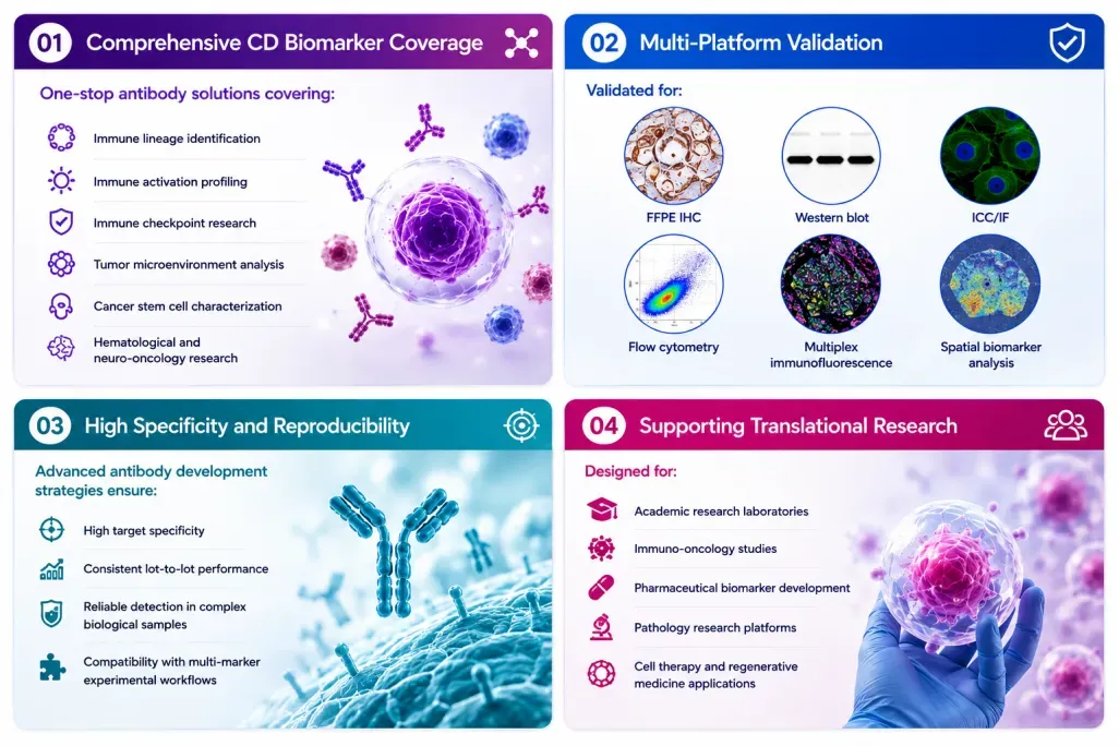
We provide comprehensive CD marker solutions enabling precise characterization of cellular identity, immune function, tumor biology, and therapeutic response. By integrating validated CD antibodies with advanced profiling technologies, researchers can define immune cell populations, analyze tumor microenvironment interactions, identify cancer stem cell populations, and investigate mechanisms of tumor progression and metastasis. Multiparametric CD marker panels further support immune activation, checkpoint pathway analysis, stem cell characterization, and spatial mapping of tissue heterogeneity. These integrated approaches accelerate discoveries in immuno-oncology, regenerative medicine, and translational biomedical research.
Why Researchers Choose Our CD Marker Solutions?
Validated Targets. Comprehensive Panels. Reliable Cellular Profiling.
Our CD antibody portfolio supports the complete cellular analysis workflow — from immune identification and tumor profiling to stem cell research and translational discovery.
Research Challenge | Our Solution |
How can I accurately identify immune cell populations? | Comprehensive lineage marker panels covering T cells, B cells, macrophages, NK cells, and dendritic cells |
How can I characterize tumor immune microenvironment? | Multiparametric CD marker combinations for immune infiltration and spatial profiling |
How can I identify cancer stem cell populations? | Validated CSC marker antibodies including CD133, CD44, CD24, CD166, and CD90 |
How can I study immune checkpoint mechanisms? | Antibodies targeting PD-1, PD-L1, CTLA-4, LAG-3, and related immune regulators |
How can I improve experimental reproducibility? | Highly validated antibodies optimized for IHC, IF, WB, and flow cytometry applications |
Key References
1. Holder A.M., Dedeilia A., Sierra-Davidson K., et al. (2024).Defining Clinically Useful Biomarkers of Immune Checkpoint Inhibitors in Solid Tumors.Nature Reviews Cancer. 24:498–512.
2. Chen J., Larsson L., Swarbrick A., et al. (2024).Spatial Landscapes of Cancers: Insights and Opportunities.Nature Reviews Clinical Oncology. 21:660–674.
3. Allam M., Coskun A.F. (2024).Combining Spatial Metabolomics and Proteomics Profiling of Single Cells.Nature Reviews Immunology. 24:701.
4. Harris M.A., Savas P., Virassamy B., et al. (2024).Towards Targeting the Breast Cancer Immune Microenvironment.Nature Reviews Cancer. 24:554–577.
5. Williams H.L., Frei A.L., Koessler T., et al. (2024).The Current Landscape of Spatial Biomarkers for Prediction of Response to Immune Checkpoint Inhibition.npj Precision Oncology. 8:178.