
info@mail.ucallm.com
From Molecular Classification to Spatial Tumor Profiling — Empowering Precision Neuro-Oncology Research with Validated Antibody Solutions
Gliomas represent the most common primary malignant tumors of the central nervous system, accounting for approximately 75–80% of malignant primary brain tumors. Characterized by remarkable molecular heterogeneity, infiltrative growth patterns, and therapeutic resistance, gliomas remain among the most challenging cancers in neuro-oncology.
Validated protein biomarkers enable accurate glioma classification, tumor grading, lineage identification, and investigation of mechanisms underlying invasion, recurrence, immune regulation, and treatment resistance.
Our Glioma Biomarker Research Solutions provide highly validated antibodies supporting comprehensive applications including:
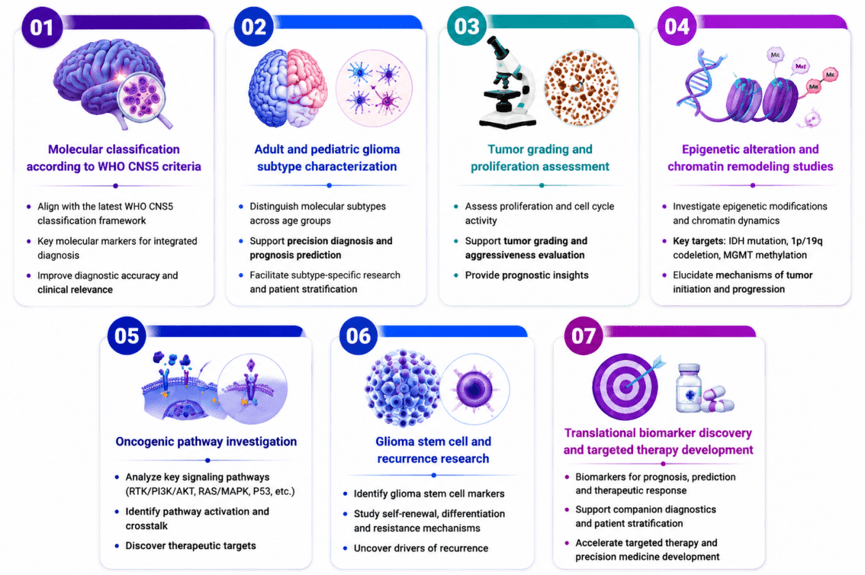
Compatible with FFPE IHC, ICC/IF and multiplex immunofluorescence, our antibody portfolio enables reliable glioma profiling from basic discovery research to translational neuro-oncology applications.
Glioma Biomarker Antibody Portfolio
——Comprehensive Solutions for Molecular Classification, Prognostic Evaluation and Mechanistic Research
Accurate glioma characterization requires integrated analysis of lineage markers, genetic alterations, epigenetic regulators, oncogenic signaling pathways, and proliferation-associated biomarkers.
Our glioma antibody portfolio covers key molecular classes defined by WHO CNS5, enabling standardized research workflows for adult and pediatric glioma studies.
Biomarker Category | Representative Markers | Research Applications | Biological Significance |
IDH-Mutant Astrocytoma Markers | IDH1 R132H, ATRX, TP53, GFAP, Ki-67,EGFR, PTEN, OLIG2 | Molecular classification, tumor grading, lineage identification | Define astrocytic lineage and molecular evolution of IDH-mutant gliomas |
Oligodendroglioma Markers | IDH1 R132H, OLIG2, TERT promoter-associated studies, CIC/FUBP1 pathway markers | Differential diagnosis and molecular typing | Support identification of IDH-mutant, 1p/19q-codeleted tumors |
IDH-Wildtype Glioblastoma Markers | EGFR, PTEN, TERT, CDKN2A/p16, Ki-67 | GBM characterization, oncogenic pathway analysis | Reflect aggressive tumor biology, proliferation and therapeutic resistance |
Diffuse Midline Glioma Markers | H3 K27M, H3K27me3 loss, EZHIP, ACVR1, TP53, OLIG2, SOX10 | Pediatric high-grade glioma diagnosis | Reveal histone-driven epigenetic dysregulation |
H3 G34-Mutant Glioma Markers | H3.3 G34R/V, ATRX, TP53, OLIG2, PDGFRA | Molecular classification and chromatin biology studies | Define hemispheric pediatric glioma driven by histone mutation |
RTK Pathway Activation Markers | EGFR, PDGFRA, MET, ALK, ROS1 | Target discovery and pathway activation studies | Identify growth signaling abnormalities driving glioma progression |
MAPK Pathway Markers | BRAF V600E, FGFR1 | Pediatric low-grade glioma research | Characterize MAPK-driven tumor development |
Proliferation and Malignancy Markers | Ki-67, PCNA | Tumor aggressiveness evaluation | Quantify proliferative activity and malignant potential |
Key Glioma Biomarker Targets
——Molecular Function and Research Applications
Marker | Biological Function | Research Value | Associated Glioma Types |
IDH1 R132H | Mutant isocitrate dehydrogenase producing oncogenic 2-hydroxyglutarate | Essential diagnostic biomarker for IDH-mutant gliomas | Astrocytoma, oligodendroglioma |
ATRX | Chromatin remodeling protein maintaining genomic stability; Loss of ATRX expression serves as a surrogate marker for ATRX mutation and alternative lengthening of telomeres (ALT). | Detect ATRX loss and distinguish astrocytoma from oligodendroglioma | IDH-mutant astrocytoma |
TP53 | Tumor suppressor regulating DNA damage response and apoptosis | Study genomic instability and malignant progression | Astrocytoma, pediatric high-grade glioma |
GFAP | Astrocytic intermediate filament protein | Identify astrocytic lineage and tumor differentiation status | Astrocytoma, glioblastoma |
EGFR | Receptor tyrosine kinase promoting proliferation and invasion | Investigate oncogenic signaling and therapeutic targets | IDH-wildtype glioblastoma |
H3K27me3 | Loss of H3K27me3 indicates PRC2 dysfunction associated with H3 K27-altered diffuse midline glioma. | Defining biomarker for diffuse midline glioma | Pediatric diffuse midline glioma |
H3.3 G34R/V | Histone mutation affecting chromatin accessibility | Characterize pediatric hemispheric glioma | H3 G34-mutant glioma |
EZHIP | Epigenetic regulator inducing PRC2 inhibition | Study H3K27me3 dysregulation | Diffuse midline glioma |
PDGFRA | Growth factor receptor driving RTK signaling | Evaluate oncogenic activation and targeted therapy opportunities | Pediatric and adult high-grade gliomas |
BRAF V600E | Constitutively active MAPK pathway driver | Identify MAPK-dependent glioma biology | Pediatric low-grade glioma |
NTRK1/2/3 | Neurotrophic receptor tyrosine kinases | Detect fusion-driven targeted therapy opportunities | Infant-type hemispheric glioma |
Ki-67 | Nuclear proliferation marker | Evaluate tumor proliferation index and malignancy grade | All glioma subtypes |
Featured Glioma Research Panels
——Integrated Biomarker Solutions for Comprehensive CNS Tumor Profiling
n Adult IDH-Mutant Glioma Classification Panel—Defining Molecular Identity of Diffuse Adult Gliomas
Key Markers
IDH1 R132H | ATRX | TP53 | CDKN2A/p16 | GFAP | Ki-67 |
n Glioblastoma Molecular Profiling Panel—Investigating Aggressive Tumor Biology and Therapeutic Resistance
Key Markers
EGFR | PTEN | TERT | p16/CDKN2A | Ki67 | TP53 |
n Pediatric High-Grade Glioma Panel—Decoding Histone Mutation-Driven Tumorigenesis
Key Markers
H3 K27M | H3K27me3 | EZHIP | ACVR1 | OLIG2 | TP53 |
n Pediatric Low-Grade Glioma Panel—Pediatric Low-Grade Glioma Panel
Key Markers
BRAF V600E | FGFR1 | MYB | MYBL1 | NF1 | CDKN2A |
Glioma Biomarker Research
——From Molecular Profiling to Translational Neuro-Oncology
We provide comprehensive glioma biomarker research solutions supporting systematic tumor characterization from molecular identity determination to disease progression analysis and therapeutic discovery. By integrating validated biomarkers for tumor lineage, molecular classification, epigenetic regulation, signaling pathways, and therapeutic vulnerabilities, researchers can establish robust frameworks for glioma diagnosis, stratification, and precision oncology research. Advanced biomarker profiling approaches further enable investigation of tumor heterogeneity, malignant progression, microenvironmental interactions, and treatment response mechanisms, supporting the discovery of clinically relevant biomarkers and novel therapeutic strategies in neuro-oncology.
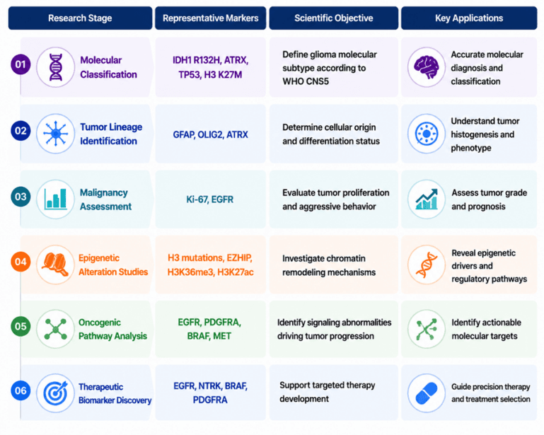
Research Frontiers Driving Glioma Biomarker Discovery
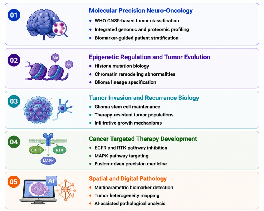
We enable comprehensive glioma research by supporting investigation across key biological frontiers, from molecular classification and tumor lineage characterization to disease evolution, therapeutic vulnerability, and spatial tumor complexity. Validated research tools facilitate the study of glioma identity, genetic and epigenetic alterations, oncogenic signaling pathways, and mechanisms underlying tumor progression and treatment resistance. Advanced multiplex profiling and spatial analysis approaches further reveal intratumoral heterogeneity, microenvironmental interactions, and cellular dynamics within the glioma ecosystem. Together, these integrated strategies empower researchers to decode glioma biology, identify disease-associated biomarkers, improve molecular stratification, and accelerate precision therapeutic discovery in neuro-oncology.
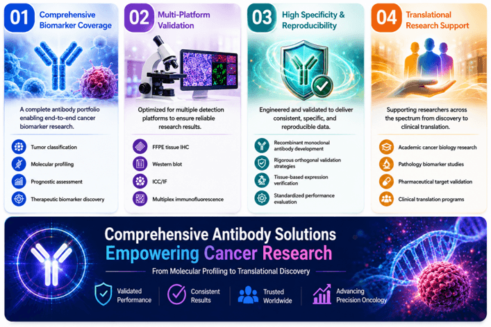
Why Researchers Choose Our Glioma Biomarker Solutions?
Validated Targets. Comprehensive Coverage. Precision CNS Tumor Research.
Our antibody portfolio supports the complete glioma research workflow—from molecular classification and pathway discovery to translational biomarker validation.
Research Challenge | Our Solution |
How can I classify glioma according to WHO CNS5 criteria? | Comprehensive molecular marker panels covering IDH1, ATRX, TP53, H3 mutations and lineage markers |
How can I distinguish glioma subtypes? | Validated antibodies for astrocytoma, oligodendroglioma, GBM and pediatric glioma classification |
kmHow can I study aggressive tumor behavior? | Biomarkers targeting proliferation, RTK signaling and invasion-associated pathways |
How can I investigate pediatric glioma biology? | Dedicated histone mutation and developmental signaling marker panels |
How can I improve experimental reproducibility? | Application-validated antibodies optimized for IHC, WB, IF and multiplex analysis |
Key References
1. Weller M., Wen P.Y., Chang S.M., et al. (2024).Glioma.Nature Reviews Disease Primers. 10:33.
2. Rudà R., Horbinski C., van den Bent M., et al. (2024).IDH inhibition in gliomas: from preclinical models to clinical trials.Nature Reviews Neurology. 20:395–407.
3. Alnahhas I. (2024).Molecular Testing in Gliomas: What is Necessary in Routine Clinical Practice?Current Oncology Reports. 26:1277–1282.
4. Taori S., Habib A., Adida S., et al. (2025).Circulating biomarkers in high-grade gliomas: current insights and future perspectives.Journal of Neuro-Oncology. 172:41–49.
5. Jones J.J., Nguyen H., Wong S.Q., et al. (2024).Plasma ctDNA liquid biopsy of IDH1, TERTp, and EGFRvIII mutations in glioma.Neuro-Oncology Advances. 6(1).