
info@mail.ucallm.com
From Immune Profiling to Precision Immuno-Oncology — Empowering Comprehensive Tumor Microenvironment Characterization with Validated Antibody Solutions
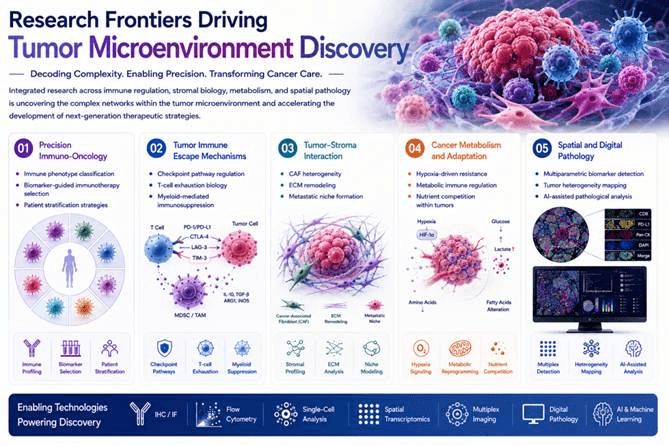
The tumor microenvironment (TME) has emerged as a central regulator of cancer progression, immune evasion, therapeutic resistance, and treatment response. Beyond serving as a structural scaffold, the TME represents a highly dynamic and complex ecosystem composed of tumor-infiltrating immune cells, cancer-associated fibroblasts (CAFs), endothelial cells, extracellular matrix (ECM), cytokines, and metabolic regulators that continuously interact with malignant cells.
We support advanced tumor microenvironment (TME) research by enabling comprehensive investigation of immune regulation, tumor–stroma interactions, metabolic adaptation, spatial heterogeneity, and therapeutic response. Validated TME biomarker solutions empower researchers to characterize immune phenotypes, investigate checkpoint regulation, T-cell exhaustion, and myeloid-mediated immunosuppression, and dissect CAF heterogeneity and ECM remodeling mechanisms underlying tumor progression and immune evasion.
Tumor Microenvironment Biomarker Antibody Portfolio
——Comprehensive Solutions for Immune Profiling, Stromal Analysis and Immunotherapy Research
Characterizing the tumor microenvironment requires integrated analysis of immune regulation, cellular composition, stromal remodeling, vascular biology, and metabolic adaptation.
Our antibody portfolio covers major TME biomarker categories to support standardized tumor immune profiling workflows.
Biomarker Category | Representative Markers | Research Applications | Biological Significance |
Immune Checkpoint Markers | PD-L1, PD-1, LAG3, TIM-3, VISTA, CTLA-4
| Immunotherapy biomarker evaluation, immune escape studies | Define immunosuppressive signaling networks and checkpoint activation status |
Immune Cell Lineage Markers | CD3, CD4, CD8, CD20, CD56 | Immune infiltration profiling and spatial immune mapping | Quantify tumor-infiltrating lymphocyte populations and immune architecture |
Macrophage & Myeloid Markers | CD68, CD163, CD206, ARG1, MPO | TAM characterization and immune polarization studies | Reveal inflammatory versus immunosuppressive myeloid states |
Stromal & CAF Markers | α-SMA, FAP, PDGFR-β, Fibronectin, Collagen I/III/IV | CAF activation and ECM remodeling studies | Characterize tumor fibrosis, matrix remodeling, and stromal regulation |
Angiogenesis Markers | CD31, CD34, VEGF-A, ANGPT2 | Tumor vascular analysis and anti-angiogenic therapy studies | Evaluate vessel formation and angiogenic signaling |
Hypoxia & Metabolic Markers | HIF-1α, CAIX, GLUT1, LDHA | Tumor stress adaptation and metabolic research | Identify hypoxic niches and metabolic reprogramming |
Cytotoxic Immune Markers | Granzyme B, Perforin, IFN-γ | Immune killing activity assessment | Evaluate functional anti-tumor immune responses |
Key Tumor Microenvironment Biomarkers
——Molecular Function and Research Applications
Marker | Biological Function | Research Value | Major Applications |
PD-L1 (CD274) | Immune checkpoint ligand expressed by tumor and immune cells | Predictive biomarker for immune checkpoint blockade response | TPS/CPS scoring, immunotherapy studies |
PD-1 | Immune inhibitory receptor on activated/exhausted T cells | Evaluates T-cell exhaustion phenotype | Tumor immune suppression analysis |
LAG3 | Co-inhibitory immune receptor | Emerging immunotherapy target with PD-1 synergy | Combination checkpoint therapy research |
TIM-3 | Exhaustion-associated receptor on T/NK cells | Identifies dysfunctional immune populations | Immune-resistant tumor profiling |
VISTA | Myeloid-associated checkpoint regulator | Characterizes myeloid-mediated immune suppression | Immunosuppressive TME studies |
CD3 | Pan-T cell marker | Measures total T-cell infiltration | Immune infiltration scoring |
CD8 | Cytotoxic T lymphocyte marker | Indicator of anti-tumor immune activity | Prognostic immune profiling |
CD20 | B-cell lineage marker | Identifies B-cell infiltration and TLS formation | Spatial immune architecture analysis |
CD68 | Pan-macrophage marker | Quantifies tumor-associated macrophages | TAM density evaluation |
CD163 | M2-like macrophage marker | Indicates immunosuppressive macrophage polarization | Tumor progression and metastasis studies |
CD206 | Alternative M2 macrophage marker | Reflects IL-10/TGF-β-associated immune regulation | Macrophage functional analysis |
α-SMA | Activated fibroblast marker | Identifies myofibroblastic CAF populations | Stromal activation studies |
FAP | CAF-specific surface protein | Defines tumor-associated fibroblast populations | CAF-targeted therapy research |
PDGFR-β | Stromal/pericyte receptor | Evaluates vascular-supporting stromal cells | Angiogenesis studies |
Fibronectin | ECM remodeling protein | Reflects invasive tumor matrix remodeling | Tumor invasion analysis |
CD31 | Endothelial cell marker | Gold-standard vascular marker | Microvessel density quantification |
VEGF-A | Major angiogenic growth factor | Measures angiogenic activity | Anti-angiogenic therapy evaluation |
HIF-1α | Hypoxia transcription factor | Identifies hypoxic tumor regions | Therapy resistance studies |
CAIX | Hypoxia-induced membrane protein | Maps oxygen-deficient tumor niches | Hypoxia biomarker analysis |
GLUT1 | Glucose transporter | Indicates increased glycolytic metabolism | Warburg effect studies |
LDHA | Lactate metabolism enzyme | Links metabolism with immune suppression | Tumor metabolic profiling |
Granzyme B | Cytotoxic effector molecule | Direct indicator of immune killing activity | Functional immune monitoring |
Perforin | Cytotoxic pore-forming protein | Confirms CTL/NK killing capability | Anti-tumor immunity evaluation |
CD4 | Helper T-cell co-receptor; orchestrates adaptive immunity. | Distinguishes T-helper subsets and their contribution to anti-tumor immunity | T-cell subset characterization, spatial immune profiling |
CD56 (NCAM1) | NK cell and NKT cell adhesion molecule. | Identifies NK cell infiltration and innate immune activity | NK cell quantification, immune architecture mapping |
MPO | Myeloperoxidase enzyme abundant in neutrophil granules. | Marker for tumor-associated neutrophils (TANs) | Myeloid cell phenotyping, inflammation studies |
ARG1 | Arginase-1 enzyme depleting L-arginine; expressed in M2 macrophages and MDSCs. | Functional marker of immunosuppressive myeloid cells | TAM/MDSC functional analysis, metabolic immune regulation |
CD34 | Sialomucin on endothelial and hematopoietic stem cells. | Microvessel density quantification alongside CD31 cells | Angiogenesis analysis, vascular niche research |
Featured Tumor Microenvironment Research Panels
——Integrated Biomarker Solutions for Comprehensive TME Profiling
Immune Checkpoint & Immunotherapy Response Panel—Immune Checkpoint & Immunotherapy Response Panel
Key Markers
PD-L1 | PD-1 | LAG3 | TIM-3 | VISTA |
Tumor-Infiltrating Immune Cell Profiling Panel—Mapping Immune Architecture Within Tumor Tissue
Key Markers
CD3 | CD4 | CD8 | CD20 | CD56 |
Tumor-Associated Macrophage & Myeloid Panel—Tumor-Associated Macrophage & Myeloid Panel
Key Markers
CD56 | CD163 | CD206 | ARG1 | MPO |
CAF & ECM Remodeling Panel—Characterizing Fibroblast Activation and Tumor Matrix Dynamics
Key Markers
α-SMA | FAP | PDGFR-β | Fibronectin | Collagen I/III/IV |
Tumor Angiogenesis & Hypoxia Panel—Understanding Vascular Adaptation and Metabolic Stress
Key Markers
CD31 | CD34 | VEGF-A | HIF-1α | CAIX | GLUT1 |
Tumor Microenvironment Research Workflow
——From Immune Profiling to Translational Immuno-Oncology Discovery
Research Stage | Representative Markers | Scientific Objective |
Immune Landscape Profiling | CD3, CD8, CD20, CD56 | Characterize immune cell composition within tumors |
Immune Checkpoint Evaluation | PD-L1, PD-1, LAG3, TIM-3 | Identify immune escape mechanisms |
Myeloid Microenvironment Analysis | CD68, CD163, CD206, ARG1 | Evaluate macrophage polarization states |
Stromal Remodeling Studies | α-SMA, FAP, Fibronectin | Investigate CAF activation and ECM regulation |
Angiogenesis Analysis | CD31, CD34, VEGF-A | Quantify tumor vascular remodeling |
Hypoxia & Metabolism Research | HIF-1α, CAIX, GLUT1, LDHA | Study metabolic adaptation and resistance mechanisms |
Immunotherapy Biomarker Validation | Multiplex TME panels | Support precision immuno-oncology research |
The Tumor Microenvironment (TME) Research Workflow integrates immune profiling, checkpoint analysis, stromal characterization, angiogenesis evaluation, and metabolic investigation to advance translational immuno-oncology research. By analyzing key biomarkers including CD3, CD8, CD20, PD-L1, PD-1, LAG3, TIM-3, CD68, CD163, FAP, α-SMA, VEGF-A, HIF-1α, and CAIX, researchers can characterize immune cell composition, investigate immune escape mechanisms, assess stromal remodeling, and define tumor adaptation strategies. This comprehensive workflow supports immunotherapy biomarker discovery, multiplex spatial profiling, and precision oncology applications by enabling deeper understanding of tumor–immune interactions and therapeutic response mechanisms.
Why Researchers Choose Our Tumor Microenvironment Research Solutions?
Validated Biomarkers. Comprehensive Panels. Reliable Immuno-Oncology Discovery.
Our antibody solutions support the complete TME research workflow—from immune profiling and stromal characterization to translational biomarker validation.
Research Challenge | Our Solution |
How can I evaluate tumor immune status? | Comprehensive immune cell and checkpoint marker panels |
How can I study immunotherapy response mechanisms? | PD-1/PD-L1 and emerging checkpoint biomarker solutions |
How can I characterize tumor-associated macrophages? | CD68/CD163/CD206/ARG1 macrophage profiling panels |
How can I analyze CAF-mediated tumor progression? | FAP, α-SMA and ECM remodeling marker solutions |
How can I improve spatial tumor analysis? | Multiplex-compatible antibodies optimized for IHC and IF |

Key References
Chen J., Larsson L., Swarbrick A., Lundeberg J. (2024). Spatial landscapes of cancers: insights and opportunities. Nature Reviews Clinical Oncology. 21:660–674.
Polak R., Zhang E.T., Kuo C.J. (2024). Cancer organoids 2.0: modelling the complexity of the tumour immune microenvironment. Nature Reviews Cancer. 24:523–539.
De Martino M., Rathmell J.C., Galluzzi L., et al. (2024). Cancer cell metabolism and antitumour immunity. Nature Reviews Immunology. 24:654–669.
Yuan S., Almagro J., Fuchs E. (2024). Beyond genetics: driving cancer with the tumour microenvironment behind the wheel. Nature Reviews Cancer. 24:274–286.